
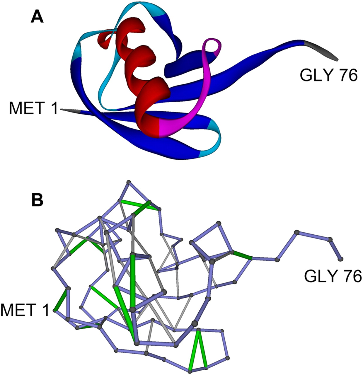
Packing deficiency of human ubiquitin. (A) Ribbon representation of ubiquitin structure (PDB. 1UBI) (Ramage et al. 1994). (B) Ubiquitin SEBH-pattern (solvent-exposed backbone hydrogen bond). The protein backbone is shown as virtual bonds (blue) joining α-carbons. Light-gray segments joining α-carbons represent well-wrapped (protected) backbone hydrogen bonds (BHBs) pairing residues defined by their α-carbon positions, and green segments represent SEBHs. The solvent-exposure extent of a hydrogen bond is determined from atomic coordinates (Fernández 2004; Pietrosemoli et al. 2007) by calculating the number of nonpolar side-chain groups within its microenvironment (see Methods). SEBHs are those BHBs protected by an insufficient number of nonpolar groups as statistically defined in Methods. The packing deficiency ν, defined as the ratio of SEBHs to the overall number of BHBs, is 31.4% (ν = 11/35).


