
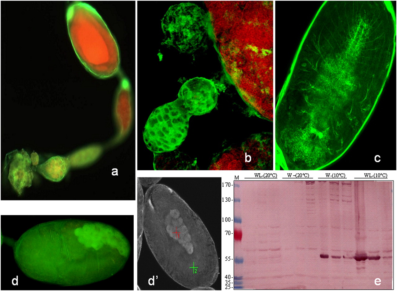
Immunohistological analysis of neuropeptides in aphid ovarioles. Dissected wingless adult ovarioles and embryos were fixed in 70% ethanol and overlaid with one of the following antibodies: anti-HRP or allatostatin followed by FITC second antibody labeling (see Supplemental material). (a) An intense fluorescent signal is evident in the ovariole envelope when stained with anti-HRP, and this pattern highlights the structural cells of the germarium (yellow). (b) Confocal microscopy shows strong fluorescent labeling of the germarium and the individualization of the first embryo. (c) Neuronal network inside an embryo labeled with an antibody anti-HRP recognizing insect neurons. (d,d′) Labeling with anti-allatostatin. The FITC fluorescent probe was confirmed by absorption spectrum in spectrometry. (e) The extracted A. pisum proteins were analyzed by a Western blot with antibodies against allatostatin (1:1000). (W) Winged aphid; (WL) wingless aphid; (M) molecular mass markers.


