
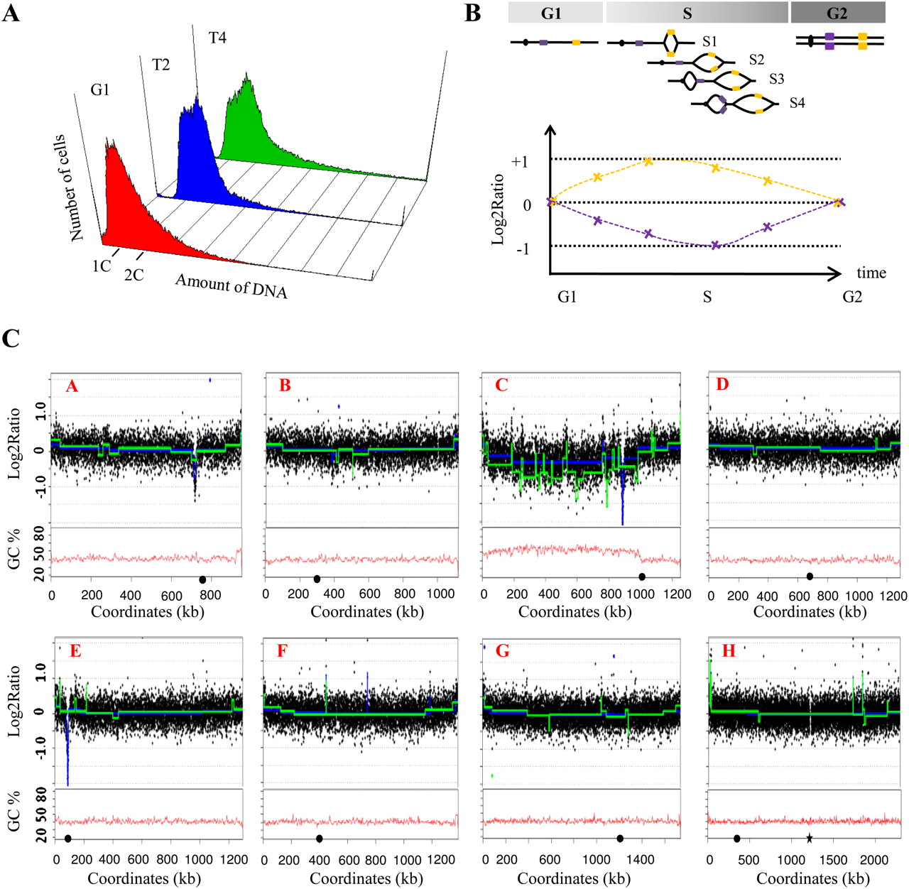
Replication timing in L. kluyveri. (A) Flow cytometry. Samples of synchronized cells were collected in G1 arrest (red) and in two points (T2 in blue and T4 in green) of the S phase spaced by 5 min. (1C, 2C) DNA content. (B) Schematic representation of the replication of a theoretical chromosome. (Yellow boxes) Early replicated regions, (violet boxes) late replicated regions. In G1, the yellow and violet regions are present in one copy. During the S phase (S1–S4), the copy number of the different cassettes increases to reach two copies in G2. The log2 ratio of the DNA content for the early (yellow) and late (violet) regions are represented according to the time from the G1 to the G2 phases. In G1, the log2 ratio is zero. At the beginning of the S phase, the regions replicated early (yellow box) are present in two copies, while the other regions are just in one copy. Thus, the log2 ratio of the yellow box tends to one, while the log2 ration of the violet box decreases. Later in the S phase, the different parts of the genome replicate until all are present in two copies, and finally in G2 the relative ratio of the intensity tends again to zero. (C) Replication profiles of the chromosomes and their GC content. Microarrays were cohybridized with the DNA from cells arrested in G1 and DNA from cells in T2 or T4 (see Methods). The ratio of the intensities of the two fluorochromes is computed and the log2 ratios are plotted according to the physical position of their corresponding sequences on the different chromosomes (A–H in red, rDNA locus ignored). The curves in blue (T2 vs. G1) and in green (T4 vs. G1) are calculated using a segmentation method for array CGH data analysis (Picard et al. 2007). The peaks underline overrepresented regions corresponding most likely to replication origins. Note that the C-left region is clearly underrepresented, suggesting that the replication of C-left is delayed compared with the rest of the genome, which is almost entirely replicated. (Black circles) Position of the centromeres, (black star) position of the rDNA.


