
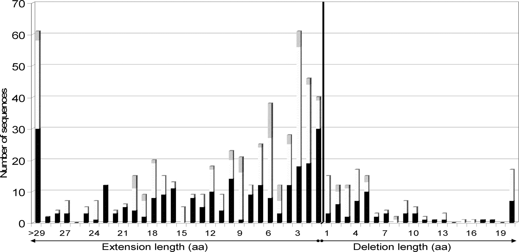
Figure 5.
Distribution of false extension and deletion lengths in mycobacteria (including M. smegmatis). Bars are shaded proportionally to the number of each actual initiation codon after correction: (black) ATG, (white) GTG, (gray) TTG.


