
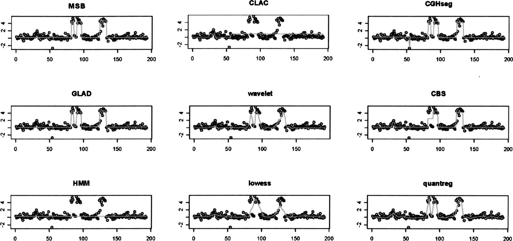
Figure 4.
Application of MSB and eight other methods to an array-CGH profile of the three amplifications around EGFR in the GBM29 sample. MSB, CGHseg, GLAD, wavelet, and quantreg clearly detected all three amplifications correctly. CBS detected three amplifications with the wrong amplitude. lowess only detected the first two amplifications as one larger region. CLAC took these three amplifications as one region. HMM performed the worst, with no detection.


