
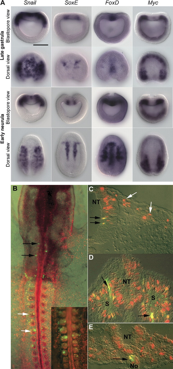
Expression of amphioxus homologues of neural crest specifier genes and reporter gene expression mediated by amphioxus FoxD (AmphiFoxD) regulatory region in chick embryos. (A) Anterior is perpendicular to the plane of the page in the blastopore view and at the top in the dorsal view. Dorsal is up in the blastopore view. Scale bar, 50 μm. Amphioxus Snail is expressed in the entire prospective neural plate and paraxial dorsal mesoderm in late gastrula stage. During the early neurula stage, Snail is down-regulated in the neural plate and transiently expressed at the neural plate border. SoxE is expressed in the dorsal mesendoderm at the border of future axial and paraxial mesoderm. FoxD is expressed in the dorsal axial mesoderm, paraxial mesoderm, and the anterior neural plate. Amphioxus Myc is expressed in the dorsal paraxial mesoderm and ventral mesendoderm. (B–E) Reporter gene expression in mesoderm derivatives and hindbrain directed by AmphiFoxD in chick embryos. Rostral is to the top in B, and dorsal to the top in C–E. EGFP+ cells have yellow or orange nuclei due to coexpression of RFP. (B) Whole-mount image of stage 11 chick embryo expressing RFP in all electroporated cells and EGFP under the control of the AmphiFoxD regulatory region; EGFP+ cells are seen in somites (white arrows, shown at higher power in inset), paraxial mesoderm, and hindbrain (black arrows). (C–E) Cross-sections showing expression of EGFP in the hindbrain (C, black arrows), somites (D, arrows), and notochord (E, arrow). No expression of EGFP is seen in premigratory or migratory neural crest cells (C, white arrows). (NT) neural tube; (S) somite; (No) notochord.


