
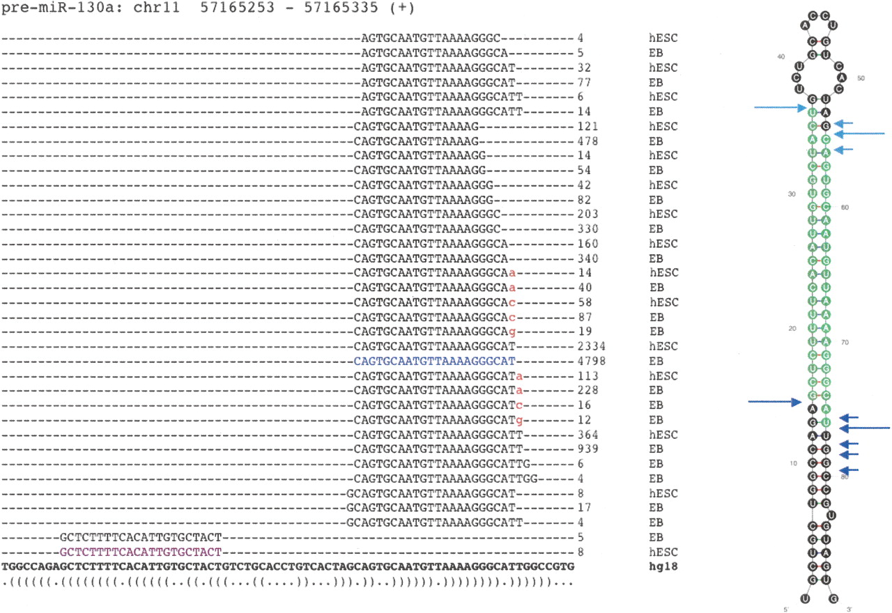
The repertoire of isomiRs and 3′ modifications of hsa-miR-130a. A diverse variety of isomiRs were observed for many of the known and novel miRNAs. Sequences representing the miR* were also commonly observed (highlighted in purple). The reference miRNA sequence from miRBase was, in many cases, not the most frequently observed isomiR (shown in blue). In this example, the most abundant miR* did not correspond to the current miRBase entry, although within the predicted pre-miRNA structure it derives from the correct 2-nt offset position relative to the reference miRNA (far right). Although variation at the 3′ end is generally much more common than at the 5′ end, target preference of the 5′ isomiRs may differ. Sequences with evidence of 3′ additions of nucleotides (red) were common, with certain miRNAs more heavily modified than others (summarized in Supplemental Table 4). The predicted structure of the pre-miRNA is represented at the bottom in dot-bracket notation, and as a graphic (far right). Experimentally inferred Drosha and Dicer1 cleavage positions are indicated in dark and light blue arrows, respectively. Large arrows represent cleavage sites for the most abundant isomiR whereas small arrows indicate those for the other isomiRs.


