
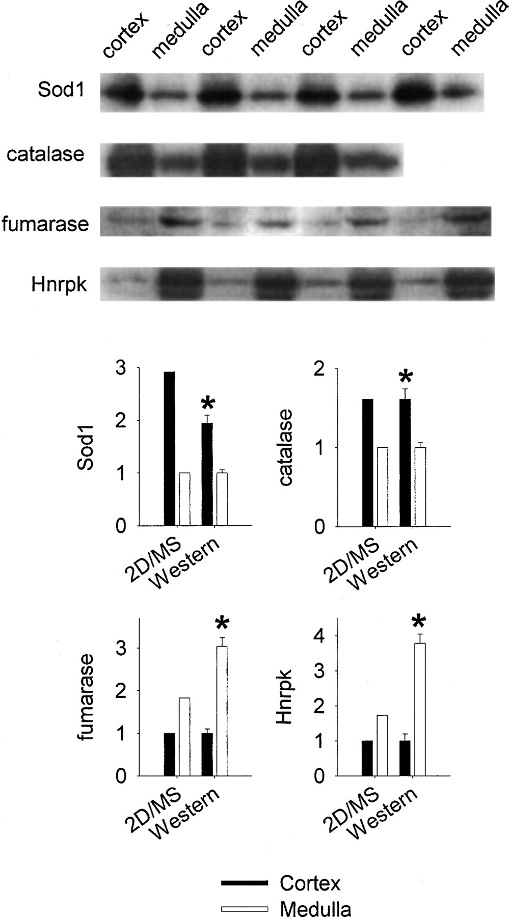
Figure 3.
Verification of protein differential expression by Western blotting. Western blots and fold differences between the renal cortex and the renal medulla measured by proteomic techniques (2D/MS) and Western blotting (Western) are shown. Band densities in Western blots were normalized to Coomassie blue staining. Note that the differences found in the proteomic analysis were statistically significant based on four rats, although error bars are not provided. (Sod1) Copper-zinc containing superoxide dismutase; (Hnrpk) heterogeneous nuclear ribonucleoprotein K. n = 3–4; (*) P < 0.05 vs. the other kidney region.


