
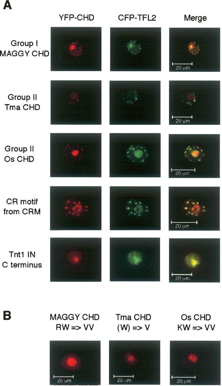
Subnuclear localization of retrotransposon CHDs and the CR motif. (A) YFP fusion proteins expressing retrotransposon CHDs or the CR motif localize to sites of heterochromatin in A. thaliana cells. Constructs expressing fusion proteins between YFP and either group I or group II CHDs or the CR motif were transformed into A. thaliana suspension cell protoplasts and visualized by confocal microscopy. The fusion proteins formed punctate foci in the nucleus and were enriched in the nucleolus. Localization was coincident with a fusion between CFP and TFL2—the A. thaliana HP1 homolog—but not the negative control, namely the Tnt1 IN C terminus. (B) Mutations in a conserved aromatic amino acid in group I and group II CHDs that is predicted to interact with the methyl group on H3 K9 abrogate subnuclear localization of YFP-CHD fusion proteins. Residues modified in the representative group I and group II CHDs are highlighted in red in Supplemental Fig. S1. In each case, residues were mutated to valine.


