
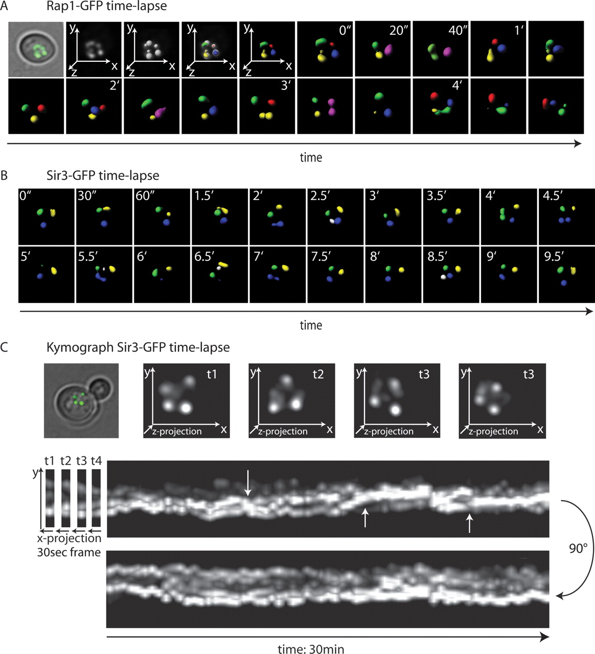
In vivo 4D fluorescence microscopy of telomeric foci. (A) Time-lapse 3D microscopy on haploid budding yeast in interphase carrying an integrated Rap1-GFP fusion under its own promoter. Microscopy was performed as described in the Methods, with stacks taken at 20-sec intervals over 90 min. Shown are 3D reconstructions from a typical 6-min series. Regions of maximal Rap1-GFP intensity were detected using the spot detection tool from the Imaris software from Bitplane, allowing each focus to be differentially labeled. When two foci fuse, they adopt one color, allowing us to score fusion and fission events. (B) Live time-lapse 3D microscopy and focus detection are as in A, but of a haploid yeast cell in interphase expressing Sir3-GFP under its own promoter. 3D stacks were taken at 30-sec intervals. (C) A kymograph of time-lapse imaging of Sir3-GFP as in B but spanning 30 min. The X-axis represents time. (Arrows) Branch or fusion points of Sir3-GFP foci. See Supplemental Movie 2 for rotation of the kymograph.


