
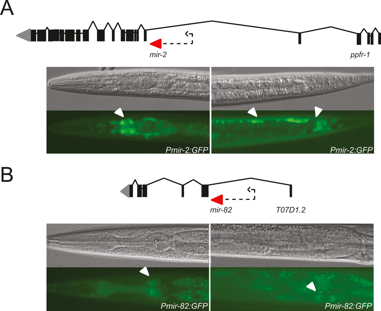
Figure 6.
Upstream sequences of two intragenic miRNAs can drive GFP expression in vivo. (A) Pmir-2 drives expression in the nerve ring (left) and ventral nerve cord and tail neurons (right). (B) Pmir-82 drives expression in pharyngeal muscle and head neurons (left), and developing spermatheca (right). (Top) DIC images; (bottom) GFP fluorescence. Arrows indicate expression. Dotted arrows indicate sequence used as promoter.


