
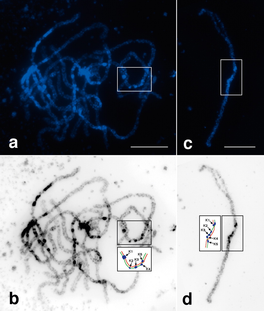
Figure 1.
Heterochromatin formation in papaya MSY. (a) A complete pachytene cell stained with DAPI. The MSY is included in the square. (b) The same pachytene cell was converted into a black-white image. The five knobs in MSY are clearly visible. The diagram in the second square illustrates the (red) X and (green) Yh chromosomes and the five (blue) knobs. (c) A complete pachytene XYh bivalent stained by DAPI. The MSY is included in the square. (d) The DAPI image was converted into a black-white image. The diagram in the second square illustrates the X (red) and Yh (green) chromosomes and the five knobs (blue). Bars, 10 μm.


