
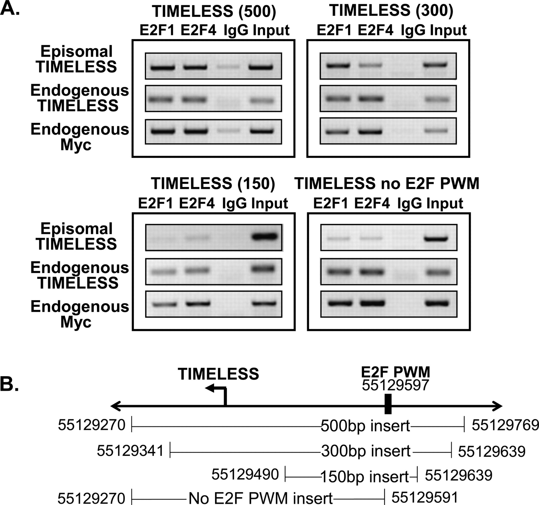
Figure 9.
Delineation of the E2F recruitment site at the TIMELESS promoter. (A) Binding of E2F1 and E2F4 to various fragments of the TIMELESS promoter is shown. The binding of E2F1 and E2F4 to the endogenous TIMELESS promoter and to the endogenous MYC promoter was also analyzed in the same samples as positive controls. (B) A schematic of the hg17 chromosomal coordinates of the tested fragments of the TIMELESS promoter is shown; the location of the best match to the E2F positional weight matrix (PWM) within the cloned in region is also indicated.


