
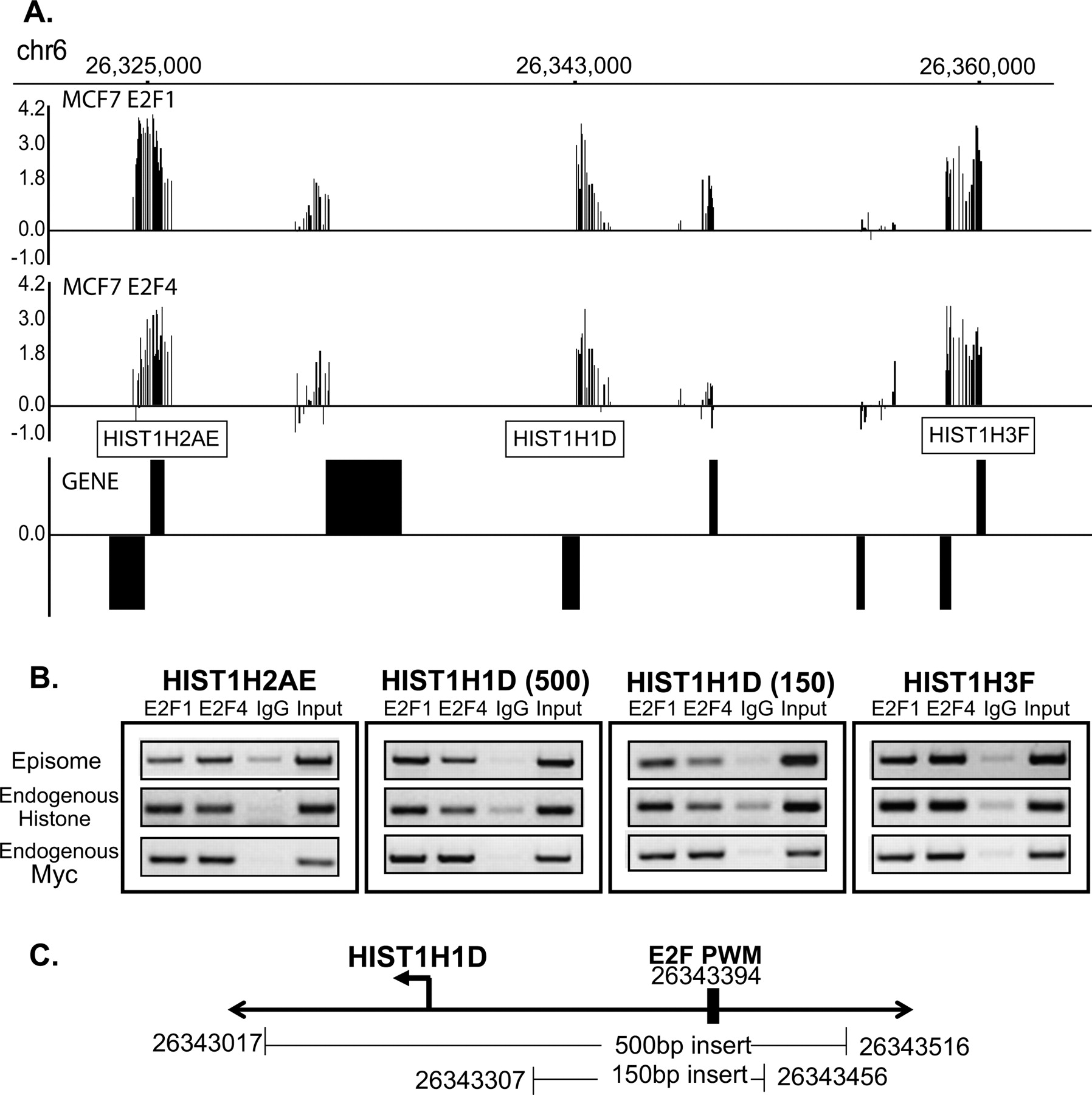
Analysis of the chromosome 6 histone cluster using eChIP assays. (A) The binding pattern of E2F1 and E2F4 in MCF7 cells to a portion of the histone cluster on chromosome 6 is shown. (B) Binding of E2F1 and E2F4 to 500-bp regions of the HIST1H2AE, HIST1H3F, and HIST1H1D promoters, as well as to a 150-bp region of the HIST1H1D promoter, is shown. The binding of E2F1 and E2F4 to the endogenous histone promoters and to the endogenous MYC promoter (as a positive control for the ChIP samples) was also analyzed. (C) A schematic of the hg17 chromosomal coordinates of the 500- and 150-bp constructs of the HIST1H1D promoter is shown; the location of the best match to E2F positional weight matrix (PWM) within the cloned in region is also indicated.


