
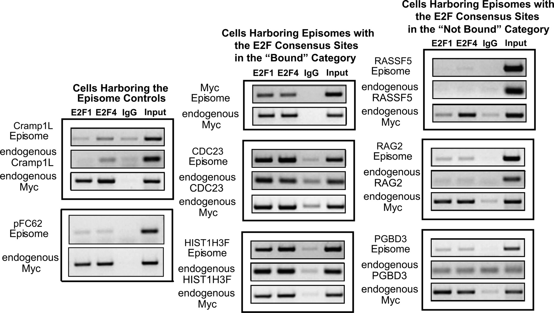
Establishment of the eChIP assay. (Left) Analysis of E2F1 and E2F4 ChIP samples in cells harboring the episomal negative controls: the empty episomal vector (left, bottom) and an episome containing a portion of the transcribed region of CRAMP1L (left, top). (Middle) Binding of E2F1 and E2F4 to episomes containing a 500-bp fragment of the MYC, CDC23, or HIST1H3F promoters, all of which possess a consensus E2F motif and were previously shown to be bound by E2Fs in the ChIP-chip experiments. (Right) Binding of E2F1 and E2F4 to episomes containing a 500-bp fragment of the RASSF5, RAG2, or PGBD3 promoters, all of which possess a consensus E2F motif, but were previously shown not to be bound by E2Fs in the ChIP-chip experiments. For all experiments, IgG was used as a negative control antibody and binding to the endogenous MYC promoter was analyzed as a positive control for E2F enrichment. Furthermore, the binding of E2F1 and E2F4 to the corresponding endogenous promoters was analyzed in all of the eChIP samples.


