
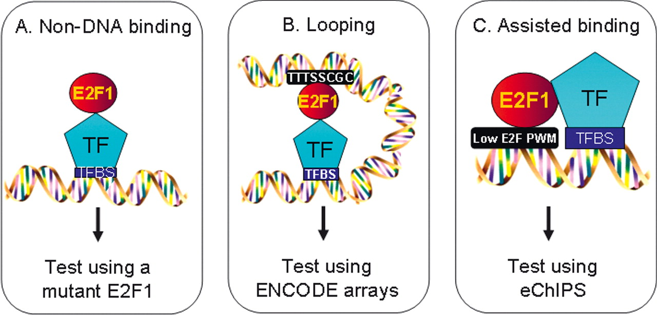
Figure 6.
Models for recruitment of E2F1 to promoters that lack a consensus motif. Shown are schematics representing potential modes of E2F1 recruitment via a mechanism independent of its DNA binding domain (A), via looping (B), or via stabilized binding in cooperation with another transcription factor (C).


