
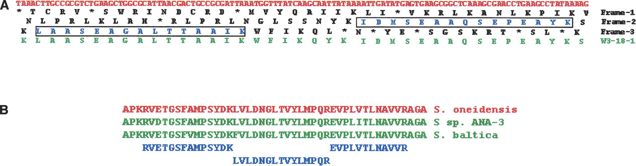
(A) The positioning of two identified peptides (boxed) relative to the three frame translation of the SO_0590 pseudogene and to homologous protein from Shewanella W3–18–1. The nucleotide sequence is shown on top (in red), and the three translated frames are shown below (in black), with stop codons translations shown as asterisk (*). The locus containing the extra A identified by re-evaluation of sequence trace TI|202865473 available in the NCBI trace archives is indicated by the arrow. (B) Alignment of identified peptides (blue) with the gene SO_4538, annotated as a degenerate M16 protease. Alignment is shown to homologous sequences in S. baltica OS155 and Shewanella sp. ANA-3.


