
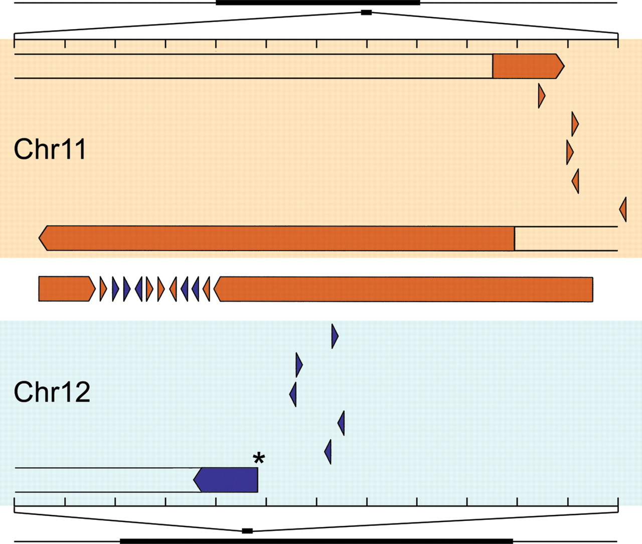
Figure 3.
Clustered genomic origins of rearranged DNA fragments. DNA segments within BAC 14g18 are shown in their reference genomic locations and orientations (see Supplemental Table 1). Fragments are shown in order 5′ to 3′ in the clone. One fragment from another BAC, 5am21, falls within the chromosome 12 interval (*). The scale shows the 120-kb region separated into 10-kb segments; the location of the expanded region is shown with respect to the amplicon for each chromosomal region. Fragments at the BAC vector insert junction are extended off-scale.


