
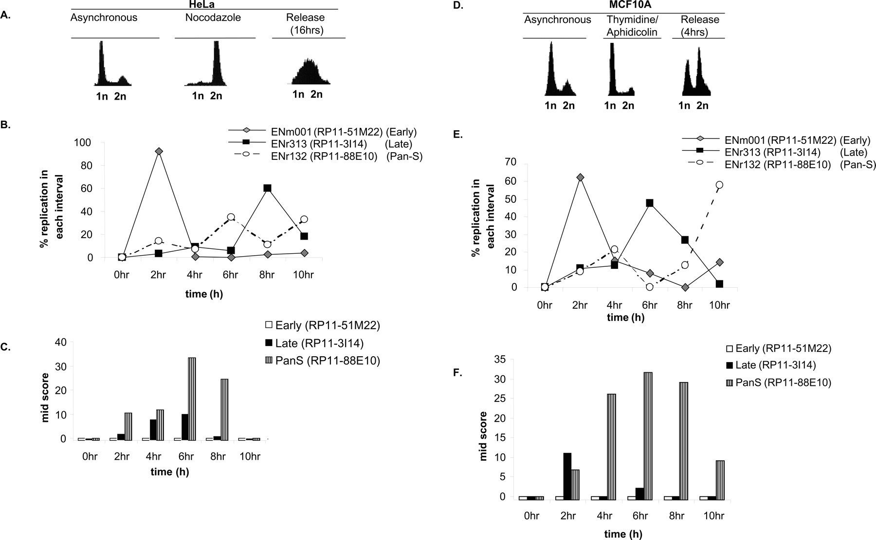
Pan-S replication pattern is independent of cell synchronization method and aneuploidy. (A) HeLa cells blocked (by nocodazole) and released from mitosis followed by FACS for DNA content. (B,C) Interphase FISH was performed with HeLa cells synchronized with nocodazole and released. The X-axis represents time in S phase such that 0 h = 12 h post-release from the nocodazole block. The rest is as in Figure 3. (D) MCF10A cells released from a G1/S block with thymidine/aphidicolin followed by FACS for DNA content. (E,F) Interphase FISH with MCF10A cells synchronously progressing through S phase to determine the replication profile and Mid-Score with the chromosomal segments mentioned in Figure 3.


