
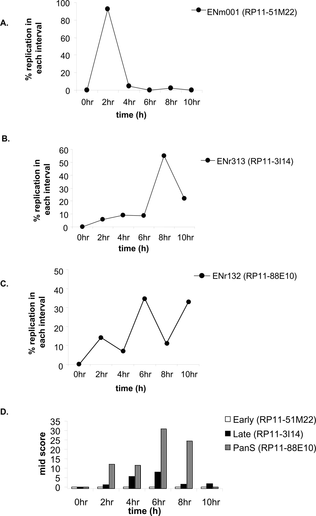
Interphase FISH for validating replication timing in HeLa. (A–C) Synchronously progressing HeLa cells were hybridized to fluorescence-labeled probes of BAC clone DNA RP11-51M22, RP11-3I14 (for early- and late-replicating areas, respectively) and RP11-88E10 (for pan-S pattern of replication). The chromosomal locations of these BACs are highlighted in Figure 2A. The percent replication at each interval of S phase is plotted against time in S phase. (D) The interallelic variation in replication for FISH data observed for each of the BAC clones mentioned above was determined by calculating the Mid-Score (detailed in Results and Supplemental Material).


