
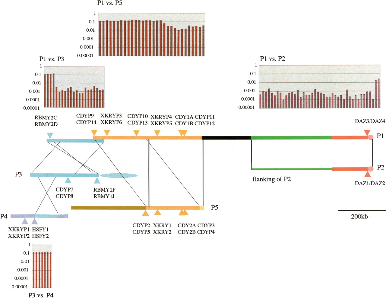
Sequence similarity of P1 against P2, P3, P4, and P5. The yellow amplicon in Kuroda-Kawaguchi et al. (2001) is divided into two: black, which has homology with autosomes, and yellow, which contains tandem duplication of a P5 ampliconic unit. Each thick line represents both arms in a palindrome, and a thin green line adjacent to P2 is nonpalindromic. Three nearly identical BYP2 copies are located in the green ampliconic unit, and two distinct pairs of PRY copies are located in a blue ampliconic unit shared by P3 and P4. In non-overlapping windows of each size 10 kb, the nucleotide differences per site (P) between P1 and other palindromic amplicons are given above P1 and below P4. Note that the P-value (ordinate) is represented in common logarithms.


