
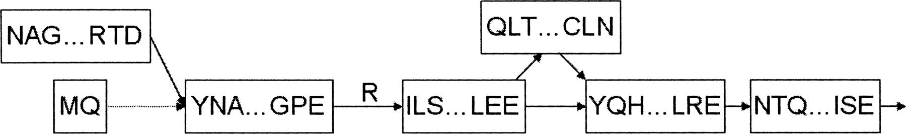
Figure 3.
A portion of the exon graph for heterogeneous nuclear ribonuclear protein K. The labeled edge represents a codon split across a splice junction. The dotted edge is an “adjacent edge” corresponding to a longer form of an exon. Searching the exon graph reveals peptides spanning both outgoing edges from the central node, confirming alternative splicing at the level of translation.


