
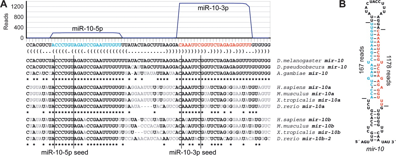
Figure 3.
Expression and conservation of mir-10. (A) The sequence and bracket-notation secondary structure of the mir-10 hairpin, highlighting the mature miR-10-5p (blue) and the mature miR-10-3p (red), with read abundance along the length of the sequence plotted above and orthologous hairpins aligned below. Nucleotides differing from the D. melanogaster identities are in gray. Vertical lines indicate the edges of the 6-nt seed of each mature RNA. (B) The mir-10 hairpin predicted secondary structure, colored as in A. Horizontal lines indicate the inferred Drosha and Dicer cleavage sites.


