
Table 2.
Percentage of repeats located at SD sequence ends
Click on table to view larger version.
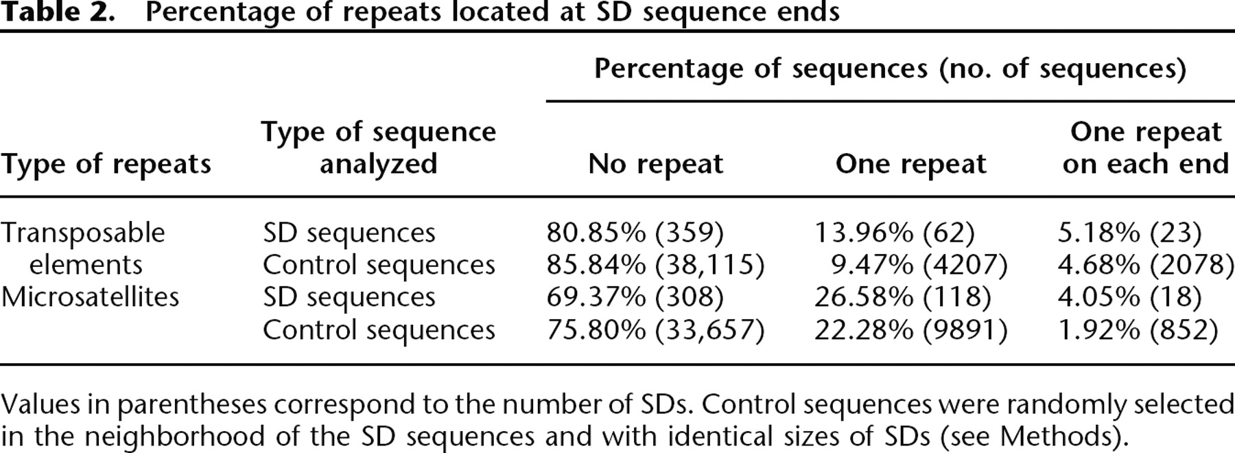
Values in parentheses correspond to the number of SDs. Control sequences were randomly selected in the neighborhood of the SD sequences and with identical sizes of SDs (see Methods).


