
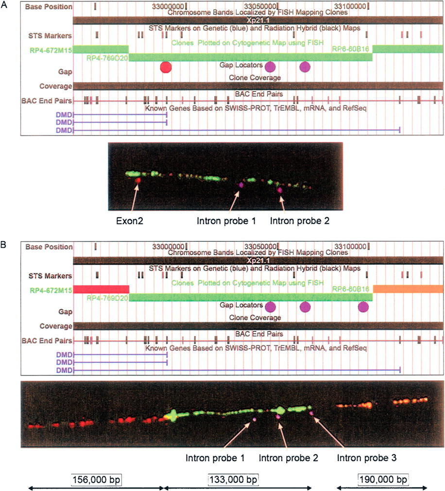
Fiber FISH image of the Dystrophin locus. Copy number variation has been identified at the Dystrophin locus in phenotypically normal humans (Iafrate et al. 2004; Conrad et al. 2006). Deletions at this locus have also been associated with Duchenne muscular dystrophy. Cytogenetic tools such as fiber FISH can be used to study the fine-scale structure of CNVs. (A) The genome structure from the UCSC genome browser showing the location of the 1-kb intron (two intron probes, purple dots), the exon (exon 2, red dot), and the three-color fiber FISH image (RP4–769D20, green). The Dystrophin locus CNV overlaps the 5′ end of Dystrophin, including exon 2 (red) and much of intron 1 (first purple dot). (B) The genome structure from the UCSC Genome Browser and the location of the 1-kb intron (three intron probes, purple dots) including non-polymorphic flanking BACs (RP4–672M15, red; RP6–60B16, orange) and a four-color fiber FISH image (RP4–769D20, green).


