
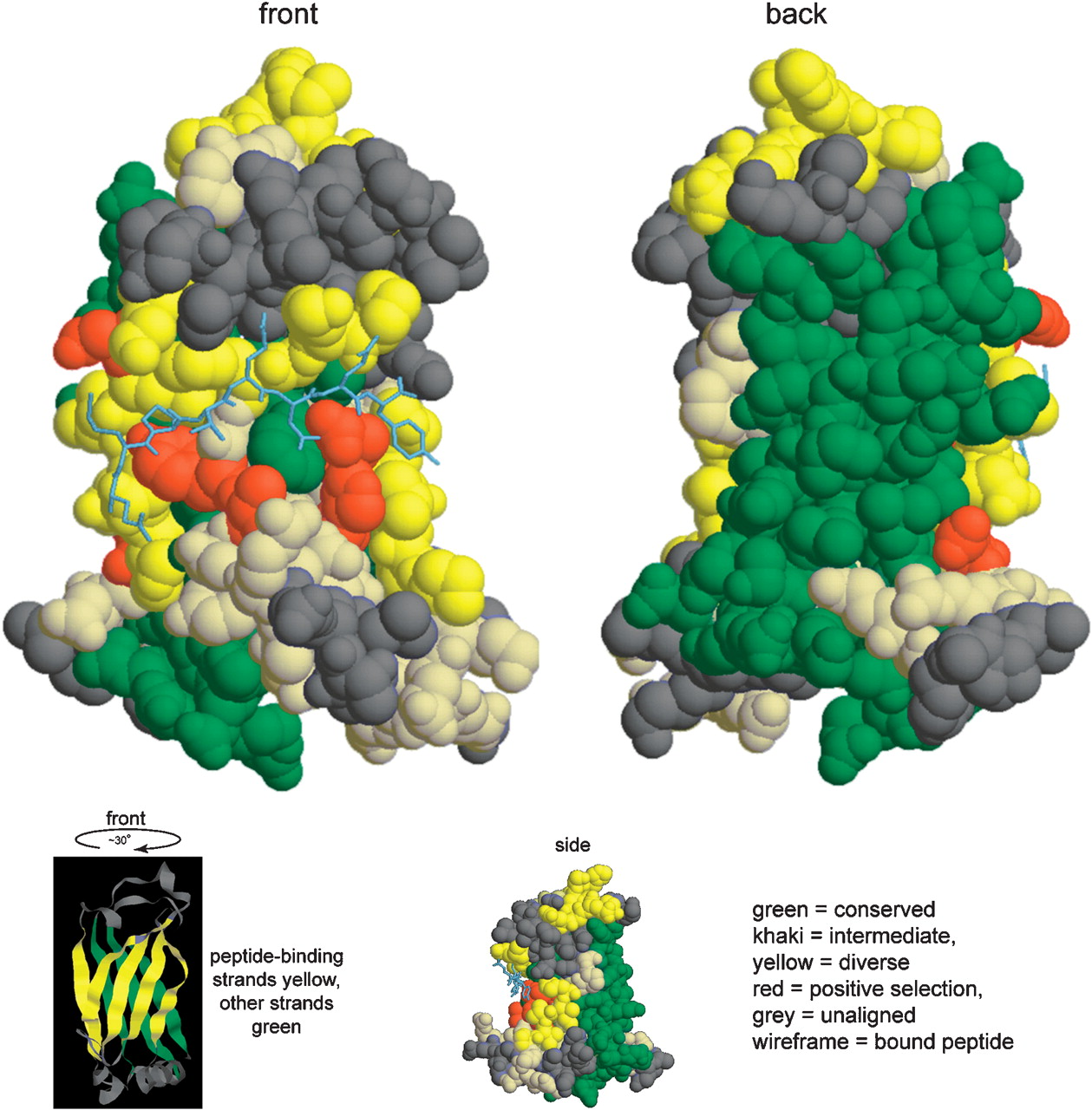
Structural model of MATH domain. The structure is TRAF6 with bound RANK peptide (PDB 1LB5), colored according to the degree of amino acid conservation among nematode MATH domains (Supplemental Fig. S11). Mapping from nematode MATH domains to TRAF6 is based on a 3D-PSSM structural alignment to the MATH domain of C08C3.2 (see Supplemental text S7). In the space-filled models, long regions of high conservation are dark green, long regions of diversity are yellow, sites of probable positive selection are red, and other regions are khaki. Residues in TRAF6 that were not aligned with C08C3.2 are gray. The bound RANK peptide is shown in gray-blue wireframe. The two large views are rotated nearly 180° from each other and are rotation-centered on the bound peptide (front) and the most conserved regions (back). The small space-filled side view shows the binding cleft in TRAF6 more clearly. The ribbon view shows the eight-stranded β-sandwich structure rotated slightly from the front view in order to show the β-strands more clearly; peptide binding strands are yellow, other strands are dark green, and non-strand regions are gray.


