
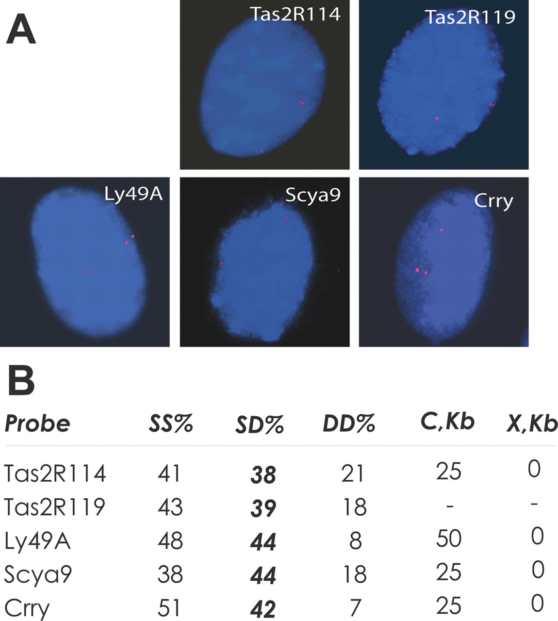
Sampling of genes in areas of tandem gene duplication reveals their asynchronous replication. (A) Mouse primary fibroblast nuclei with singlet–double (SD) hybridization pattern are shown. Red depicts PCR-generated Cy3-labeled probes for the denoted gene; blue depicts DAPI. (Supplemental Fig. S3A shows a high resolution version of this figure.) (B) Summary of the FISH assay for asynchronous replication in primary mouse fibroblasts and summary of spatial relationships (defined by the parameters C and X; see also Supplemental Fig. S3B) of the analyzed genes with clusters of related genes. (SS) Percentage of BrdU-positive nuclei with single–single hybridization signal; (SD) same for SD signal; (DD) same for double–double signal. In each case, 80–110 BrdU-positive nuclei were counted.


