
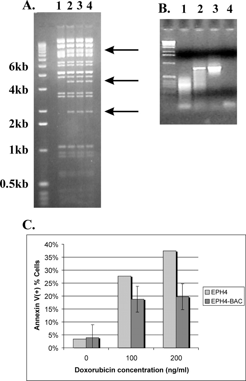
Figure 2.
Using tumor-derived BAC clones for phenotype screens. (A) Confirmation of retrofitting BAC MCF-7_1-3F5 for transfection studies. Arrows mark new bands in the vector resulting from insertion of pRetroES plasmid. (B) PCR-based control for transfection of EPH4 cells with retrofitted BAC clone. (Lane 1) Nontransfected cells; (lane 2) transfected cells; (lane 3) positive control (BAC DNA); (lane 4) negative control. (C) Increase in resistance of EPH4 cells to doxorubicin. Note the decrease in the number of apoptotic cells as demonstrated by the annexin assay.


