
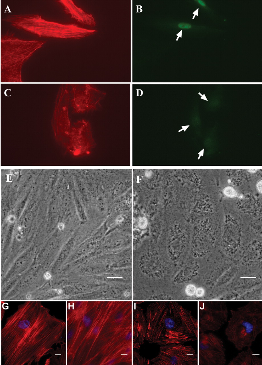
The actin cytoskeleton is dependent on SRF. (A,B,C,D) Human umbilical vein endothelial cells virally transduced for 5 d with either (A,B) shEGFP or (C,D) shSRF and then stained with phalloidin for (A,C) actin cytoskeleton or (B,D) a fluorescently tagged antibody to SRF. Arrows indicate nuclear staining for SRF. (E,F) Phase contrast micrographs of rat A7r5 smooth muscle cells transduced with (E) shEGFP or (F) shSRF for 3 d. Note the loss of cell definition in shSRF-transduced cells. This change is readily apparent by this time and remained apparent as long as 7 d post-transduction (not shown); size bars, 20 μm. (G,H) Normal cytoskeleton in A7r5 cells transduced with shEGFP for 3 and 5 d, respectively. As with human endothelial cells above, shSRF results in an alteration in normal cytoarchitecture (I) 3 d and (J) 5 d post-transduction. Note the shorter filament length, altered filament orientation, and overall lower phalloidin staining intensity in Ad-shSRF cells as compared to controls. The microtubule network in both shSRF and shEGFP transduced cells was similar, indicating the effect of shSRF is specific to the actin cytoskeleton (data not shown). Size bars, 10 μm.


