
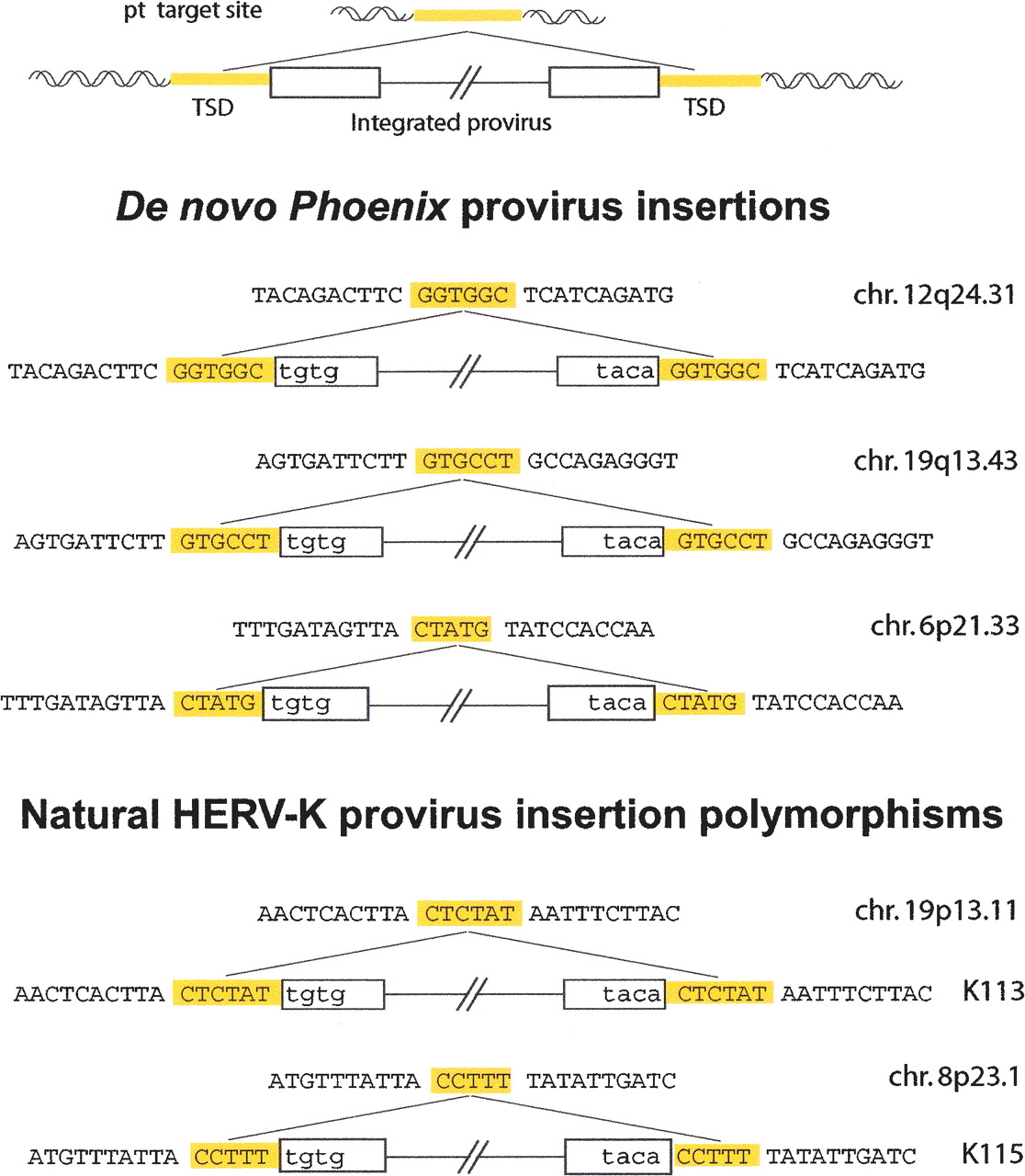
Structure of three de novo integration sites of Phoenix provirus, and comparison with the structure of natural HERV-K provirus insertions. The complete characterization of inserted Phoenix elements and insertion sites was performed using individual clones from human SH-SY5Y cells after infection and G418 selection. A provirus insertion and the corresponding empty site are schematized at the top. The sequences of three characterized Phoenix de novo insertions are shown below, with the flanking DNA in uppercase and the proviral sequences in lowercase; target-site duplications of 5/6 bp (TSD, yellow) are found in all cases, associated with full-length LTRs. For comparison, the corresponding structures of two resident HERV-K proviruses with a polymorphic insertion in humans (Turner et al. 2001) are presented in the lower part, one with a 5-bp TSD and one with a 6-bp TSD.


