
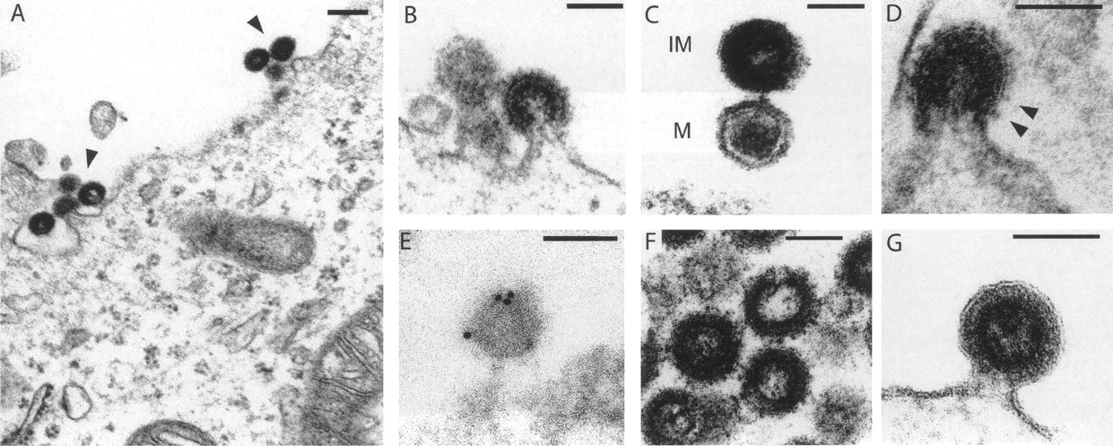
Electron microscopy of the viral-like particles generated by the Phoenix provirus. Human 293T cells were transfected with an expression vector for Phoenix (A–E), or mutants (F,G), and observed 48 h post-transfection. (A) Low magnification of particles assembled at the cell membrane. (B) Representative image of particles budding from the plasma membrane. (C) High magnification of two particles, one of which (bottom) discloses a mature (M) morphology with a condensed core, while the other appears to be still immature (IM) with two dark peripheral rings surrounding an electron-lucent core. (D) High magnification of a particle with prominent spikes, corresponding to the Env protein. (E) Image of a particle after labeling with an antibody specific for the HERV-K envelope protein and a secondary antibody linked to gold beads, obtained by immuno-electron microscopy. Quantification of the labeling on 11 independent fields demonstrates association of the gold beads with the viral particles: 307 ± 121 gold beads/μm2 for the viral particles, versus 4.9 ± 3.2 and 1.1 ± 1.5 gold beads/μm2 for the cytoplasm and particle-free extracellular space, respectively (P < 0.001 between viral particles and any of the two other compartments, Student’s t-test). (F) Image of representative particles obtained after transfection with an expression vector for the Phoenix pro mutant. All of them disclosed an immature morphology (41 of 41 identified “free” particles, i.e., no more in the budding process, for the pro mutant, vs. 15 of 37 for Phoenix WT). (G) High magnification of a particle obtained after transfection with an expression vector for the Phoenix env mutant. The membrane surrounding the particle is clearly detectable, without any spike. Scale bars: (A): 200 nm, (B–G):100 nm.


