
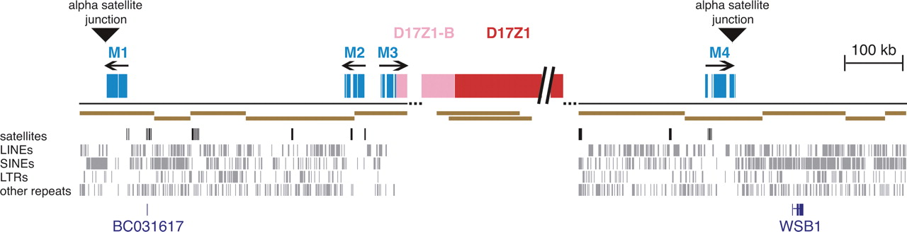
α-Satellite organization in the centromeric region of chromosome 17. The genomic landscape 500 kb distal of both sides of the centromere gap (dotted lines) is depicted. Blocks of monomeric α-satellite (light blue) are shown on both p and q arm contigs, and the p arm contig terminates with D17Z1-B higher-order α-satellite (pink). The proposed organization of D17Z1 (red) and D17Z1-B (pink) is shown inside the centromere gap. Arrows indicate the orientation of α-satellite monomers, and triangles show the junctions between α-satellite and non-satellite sequences (α-satellite junctions). BACs comprising the minimal tiling path are shown in brown, as are the two BACs containing D17Z1 at one end and D17Z1-B at the other. Other repeats are shown below the BAC contigs; from top to bottom, satellites (black), LINEs, SINEs, LTRs and other repeats (gray). The locations of RefSeq genes BC031617 and WSB1 are shown in dark blue at the bottom.


