
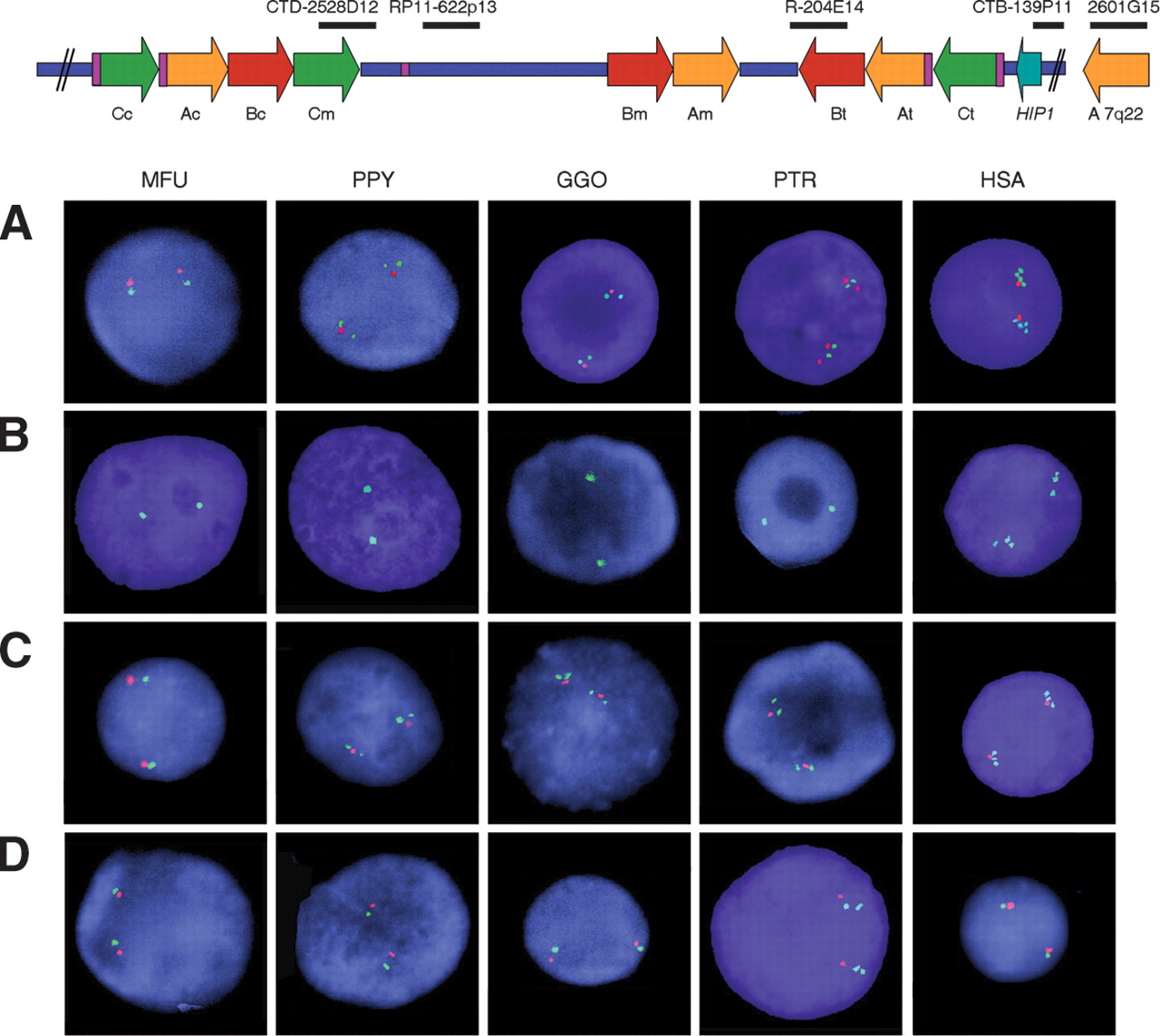
Number of blocks of segmental duplications detected by interphase FISH in the different primates with selected human BAC clones as probes. The location of all probes with respect to a representation of the human map is shown on top. (A) One single signal per chromosome in MFU, two in PPY, GGO, and PTR and four in HSA are detected with CITBI-E1-2601G15 (block A, 7q22, green), whereas CTB-139P11 (HIP1 locus, red) is single-copy in all species except chimpanzee, where it is duplicated. (B) In each species BAC RP11-204E14 (block B, green) displays one signal per chromosome except for humans, where three signals are found. (C) A single signal per chromosome is found in all the nuclei with RP11-622P13 (STX1A locus, red), whereas CTD-2528D12 (block C, green) displays one signal per chromosome in MFU, but two in PPY, GGO, and PTR and three in HSA. The single-copy STX1A locus is located in between the two (hominoids) or three (humans) signals of block C sequences. (D) CTB-139P11 (HIP1 locus, green) shows one signal per chromosome in all interphase nuclei except for PTR, which shows two signals indicating a duplication. Both copies of the HIP1 locus are located telomeric to the STX1A locus (RP11-622P13, red).


