
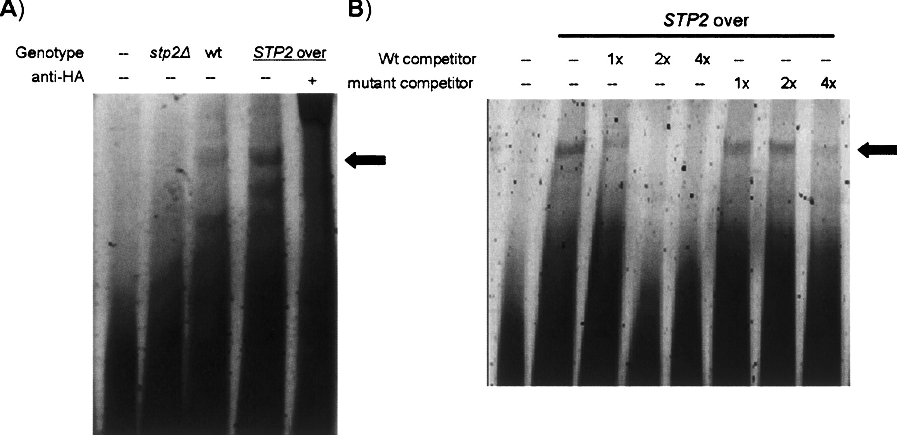
Figure 4.
Gel shift assays. (A) Gel shift showing the Cy3 labeled probe incubated with whole-cell extracts from stp2Δ cells, wild-type cells, or cells engineered to overexpress HA-tagged STP2. The black arrow represents the reproducible shift involving Stp2. Faster running bands represent possible cleavage or degradation products of Stp2. The last lane shows a super-shift with anti-HA antibody. (B) Gel shift showing STP2 overexpressing whole-cell extract with varying amounts of unlabeled wild type and mutant competitor.


