
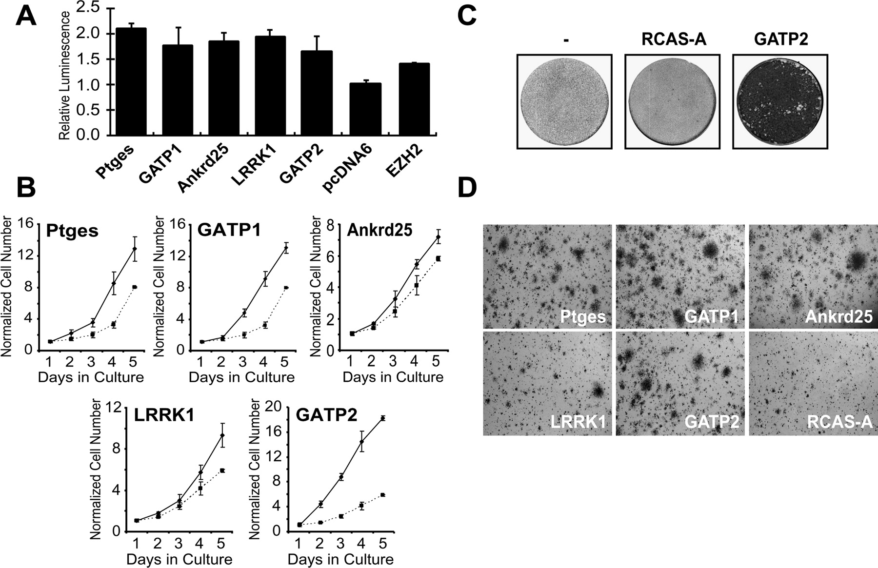
Validation of growth-activating and transforming abilities of identified proteins. (A) Overexpression of LRRK1, Ankrd25, Gatp1, Gatp2, and Ptges in HEK293 cells induces cell proliferation under serum-deprived conditions. Changes in luminescence, indicative of intrawell ATP levels, are presented relative to pcDNA6-transfected control. (B) Ectopic LRRK1, Ankrd25, Gatp1, Gatp2, and Ptges expression (solid lines, respectively) confers increased growth kinetics in primary cells, as compared to an empty vector control (dotted lines). Primary CEFs expressing growth activators were seeded into 384-well plates at a density of 500 cells per well. Cell proliferation was monitored in culture for 5 d by quantitative fluorescence imaging with the Q3DM EIDAQ100. Images were analyzed using Cytoshop, and cell numbers were determined through Hoechst 33342 staining. (C) CEFs infected with Gatp2-expressing RCAS vector grown under serum rich agar show increased saturation density compared to uninfected (–) or vector-infected (RCAS-A) controls. CEF monolayers were infected with activator-expressing virus, or the parental RCASA control virus, and subsequently overlaid with agar medium (Bister et al. 1977; Bos et al. 1990). Foci were visualized after 3–4 wk in culture by crystal violet staining. (D) CEFs ectopically expressing LRRK1, Ankrd25, Gatp1, Gatp2, and Ptges were embedded in soft agar, and colony growth was monitored over 4 wk (Bister et al. 1977). Infection with the parental viral vector RCAS-A was used as a negative control.


