
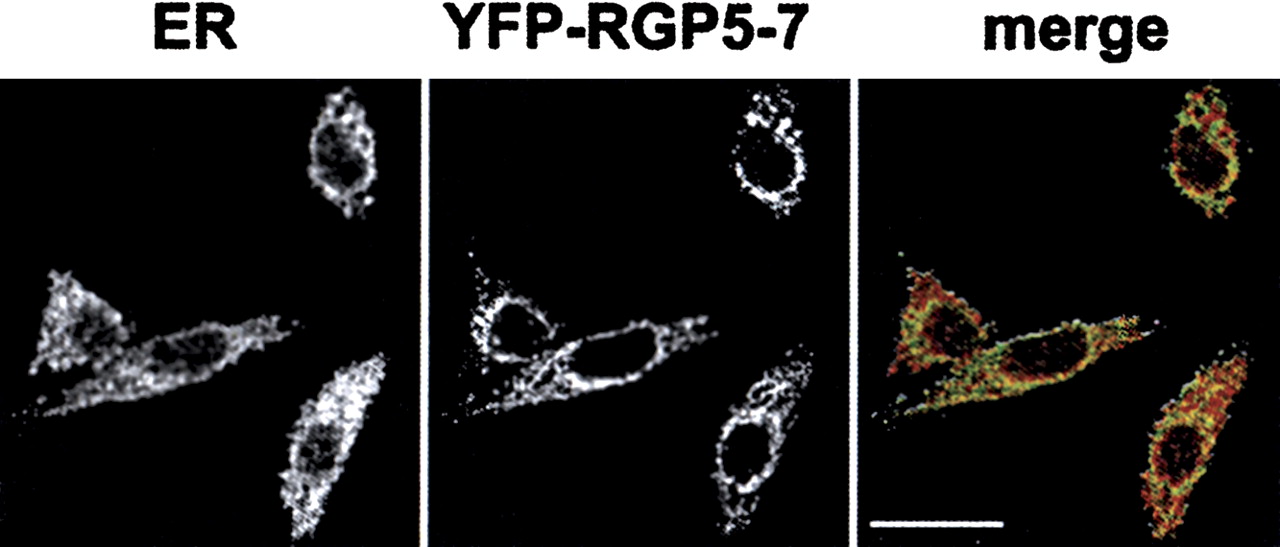
Figure 4.
Subcellular localization of RanBP2L1 isoform 2 (RGP5-7). Confocal images of fixed HeLa cells expressing a GFP-fusion of RANBPL1 isoform 2. Cells were stained with a polyclonal anti-calnexin antibody. In the merged image the calnexin signal is shown in red and the GFP signal is shown in green. Scale bar, 20 μm.


