
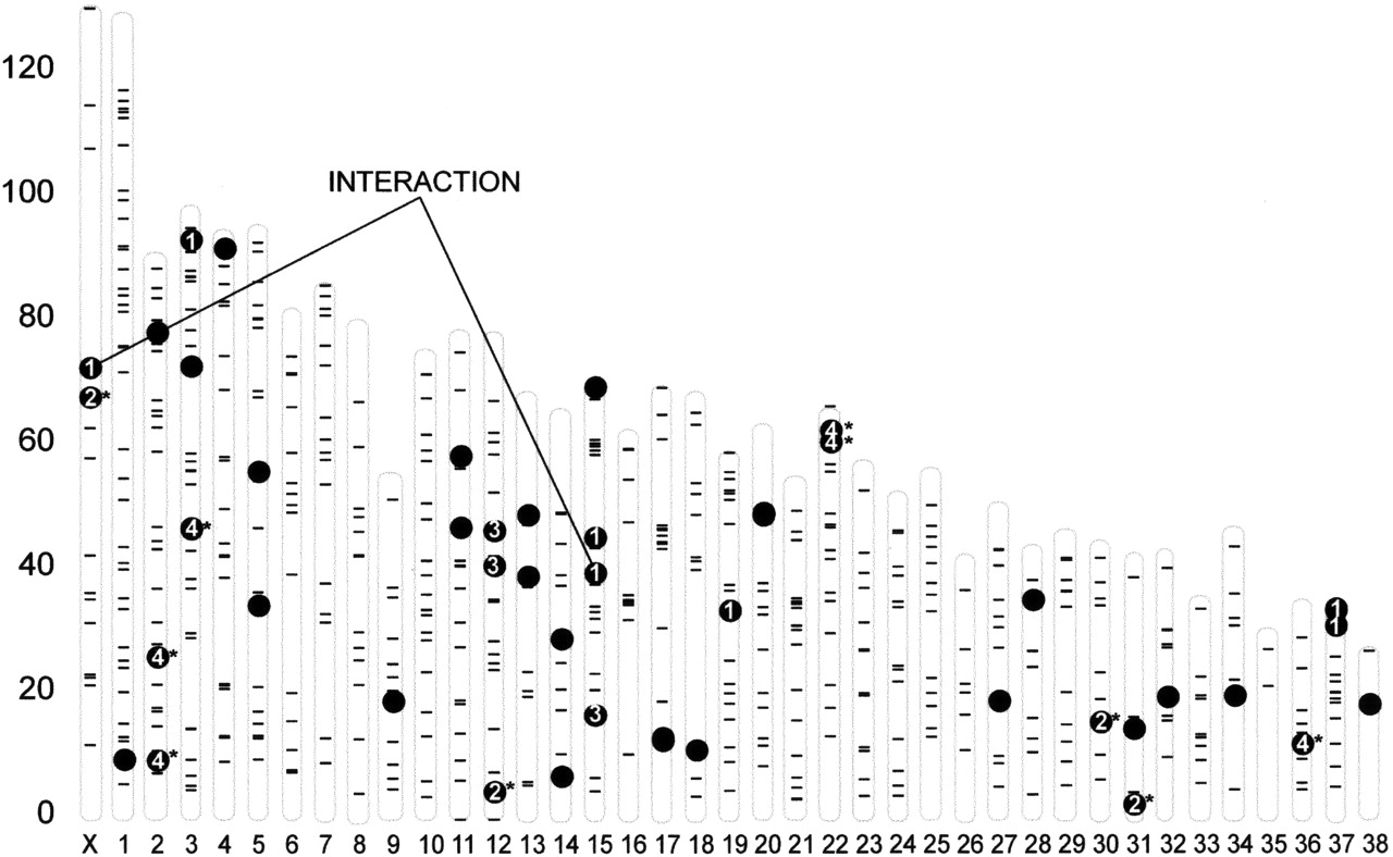
Figure 3.
Physical map of SSR markers used in QTL identification: Location of skeletal QTLs on the dog genome. For each autosome and the X chromosome, the physical positions of the markers used in the genome scan are indicated (–). Markers that show a significant association with a phenotype are indicated with black dots; those for PC1—4 are numbered. Other QTLs for PCs with significant heritabilities are indicated by additional black dots. An interaction of PC1 QTLs between the X chromosome and chromosome 15 is indicated (Chase et al. 2005b).


