
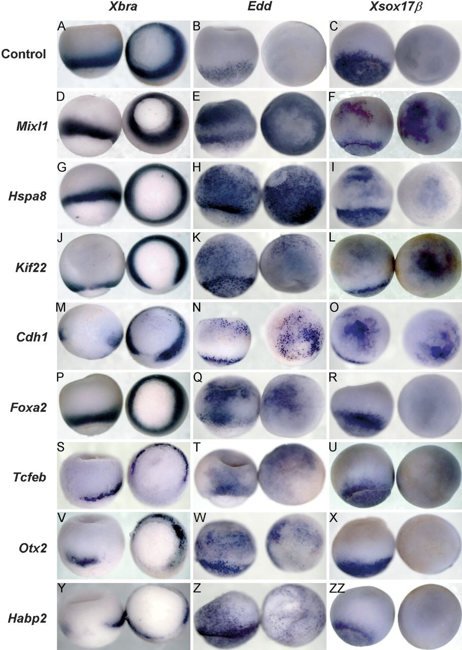
Positive clones that cause ectopic formation of endoderm or mesoderm in whole embryos. Gastrulating Xenopus embryos expressing positive clones were analyzed for the expression of Xbra (A,D,G,J,M,P,S,V,Y), Edd (B,E,H,K,N,Q,T,W,Z), or Xsox17β (C,F,I,L,O, R,U,X,ZZ). Identities of clones are listed at left and include Mixl1 (D-F), Hspa8 (G-I), Kif22 (J-L), Fzr1/Cdh1 (M-O), Foxa2 (P-R), Tcfeb (S-U), Otx2 (V-X), and Habp2 (Y-ZZ). (A-C) Uninjected controls. Concentrations of mRNAs injected into embryos are as follows: Mixl1 (D,F: 1 ng; E, 333 pg), Hspa8 (G-I: 1 ng), Kif22 (J,L: 200 pg; K, 100 pg), Fzr1 (M-O: 200 pg), Foxa2 (P-R: 200 pg), Tcfeb (S: 600 pg; T,U: 1 ng), Otx2 (V: 500 pg; W: 1 ng; X: 200 pg), Habp2 (Y: 200 pg; Z: 1 ng; ZZ: 400 pg). Embryos are oriented in Xbra column with left embryo vegetal hemisphere (endoderm) facing the bottom of the page, and right embryo vegetal hemisphere (endoderm) facing out. Embryos are oriented in both Edd and Xsox17β columns with left embryo vegetal hemisphere (endoderm) facing the bottom of the page and with the right embryo animal hemisphere (ectoderm) facing out.


