
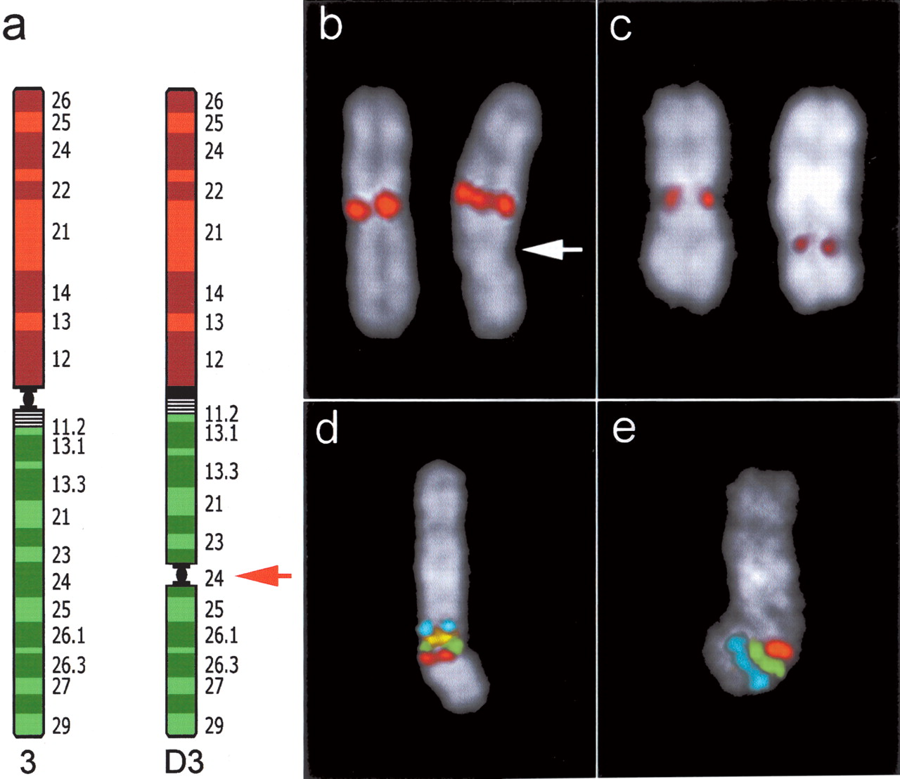
Neocentromere case 2. (a) Diagram of chromosome 3 showing the position of the neocentromere in the derivative chromosome (D3). (b) FISH signals on normal (left) and abnormal chromosome 3 (right) using an alphoid probe specific for chromosome 3. Arrow points to the neocentromere. (c) Immunotyping experiment using CREST antibodies. Signals are present at the centromere of normal chromosome 3 (left) and on the neocentromere (right). (d,e) Cohybridization experiments performed to show normal marker arrangement around the neocentromeric region. (d) FISH signals of probes RP11-21N8 (blue); RP11-220J13 (yellow); RP11-383G6 (green); RP11-505J9 (red). (e) Signals of probes RP11-505J9 (red); RP11-426N12 (green); RP11-203L15 (blue). RP11-505J9 (red, K in Fig. 1), present in both experiments, is the most (neo) centromeric probe (see text). Both experiments indicate that marker order around the neocentromere is perfectly conserved. The position of each of these BACs in UCSC database is reported in Supplemental Table 2.


