
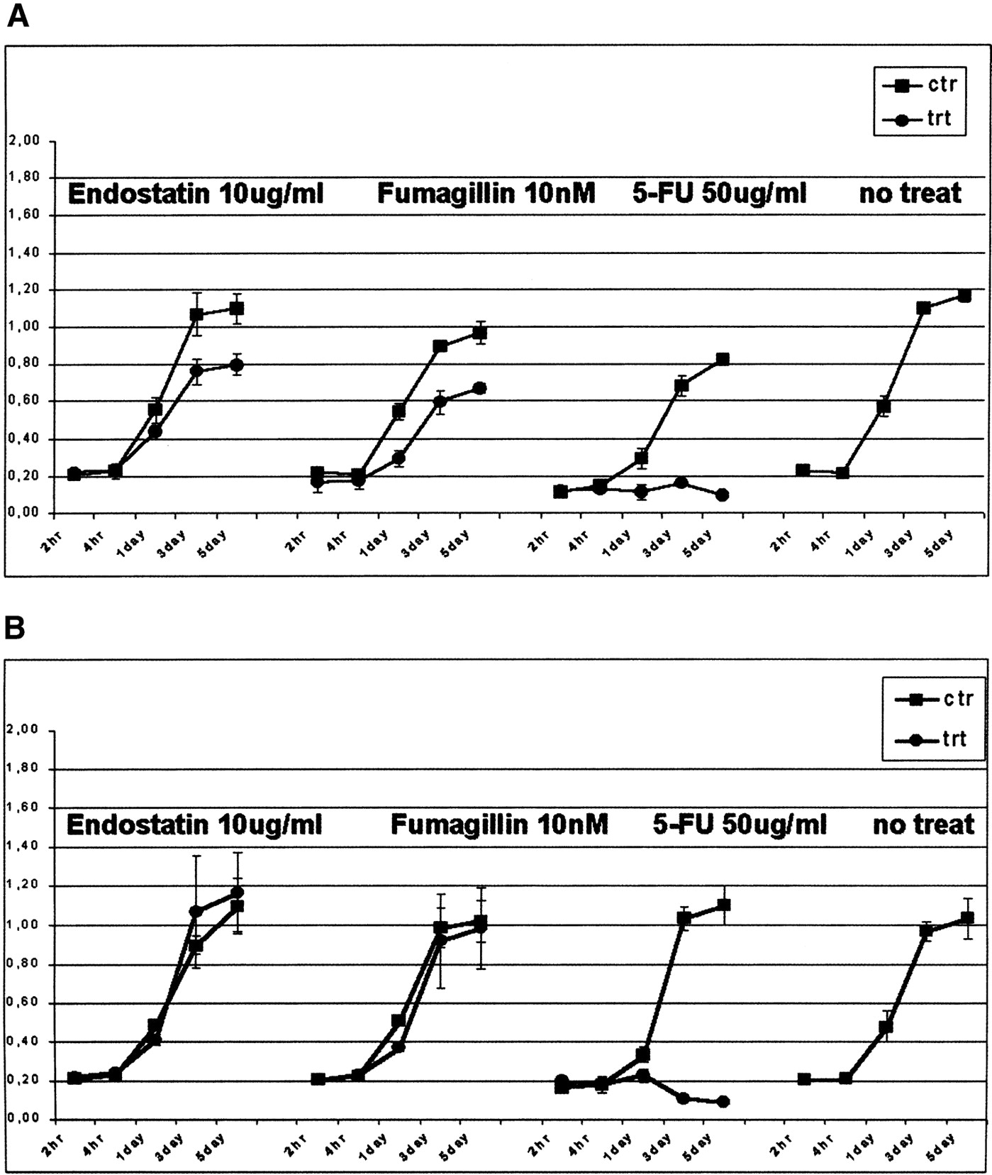
Effects of different antiangiogenic agents on cell proliferation using WST1 assay for HUVECs (A) and human fibroblasts (B). Between 2 and 4 × 10³ P2-P3 cells were plated in a 96-well tray, and treated for 2–4 h, and then 1, 3, and 5 d with 10 μg/mL endostatin, 10 nM fumagillin, and 50 μg/mL 5-FU. In A, it is possible to see a significant difference in proliferation between the treated and nontreated cells for all antiangiogenic agents at day 1. The black line indicates the proliferation rate of cells treated with medium and carrier (DMSO for fumagillin and 5FU, a citrate-phosphate buffer for endostatin, see Methods). The gray lines represent the proliferation rate of cells treated with each different reagent. The line on the far right represents cells growing in medium alone. Each data point represents the average of three experiments. The values on the y-axis are the actual WST1 fluorescent intensity readings. Endostatin and fumagillin specifically inhibited the proliferation of endothelial cells, whereas 5FU was cytotoxic for both cell types.


