
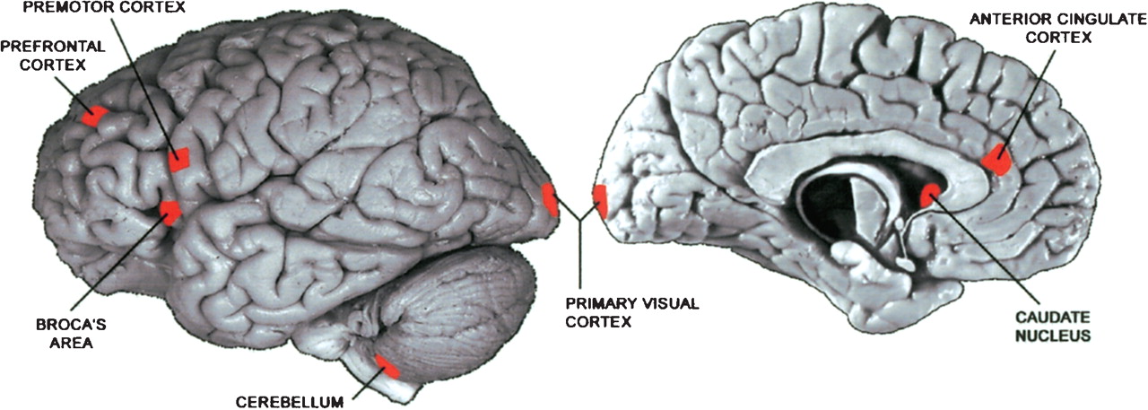
Figure 1
Location of areas sampled from the human cerebral cortex. The size of the marked areas corresponds approximately to the size of the dissected tissue sample. The sample from the right hemisphere (not shown) was taken from the location that mirrors the location of Broca's area. The human brain pictures are reprinted with permission from the Digital Anatomist Project, Department of Biological Structure, University of Washington © 1998 (http://www9.biostr.washington.edu/da.html).


