
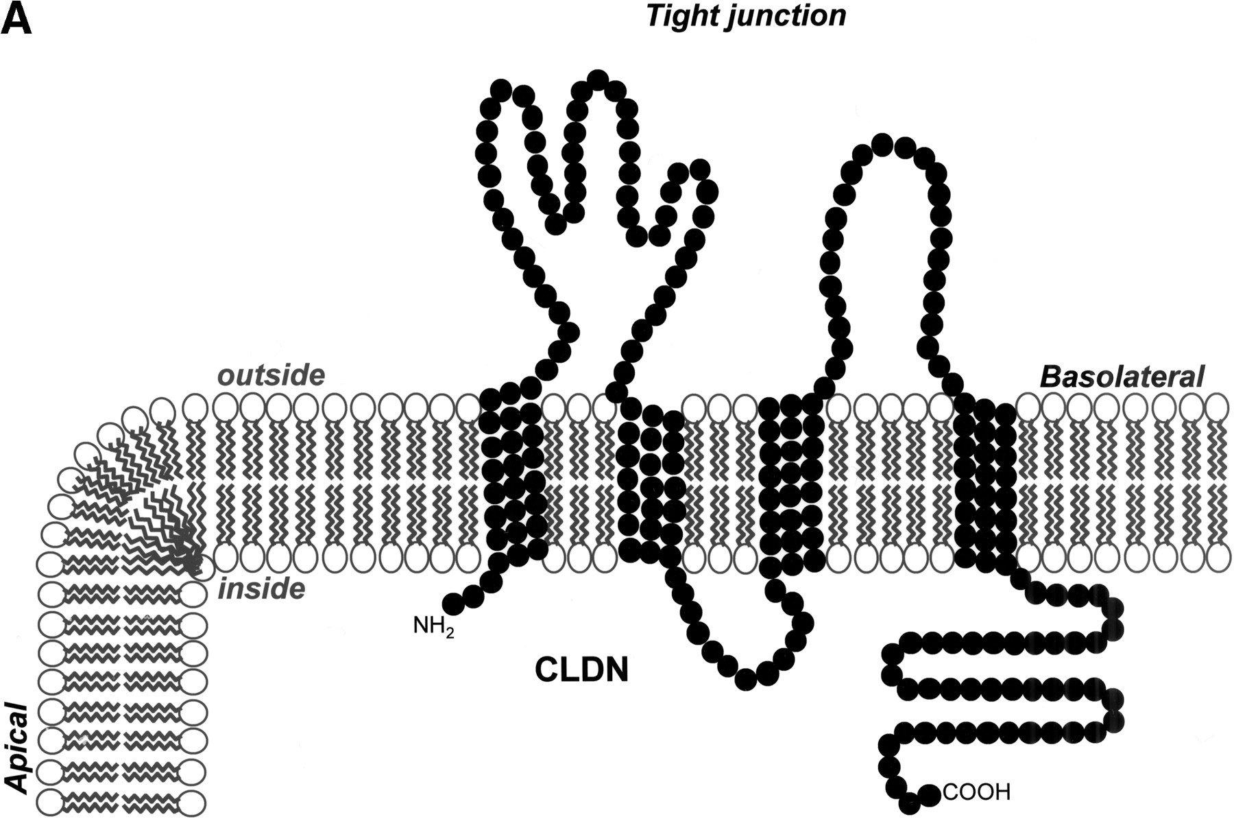
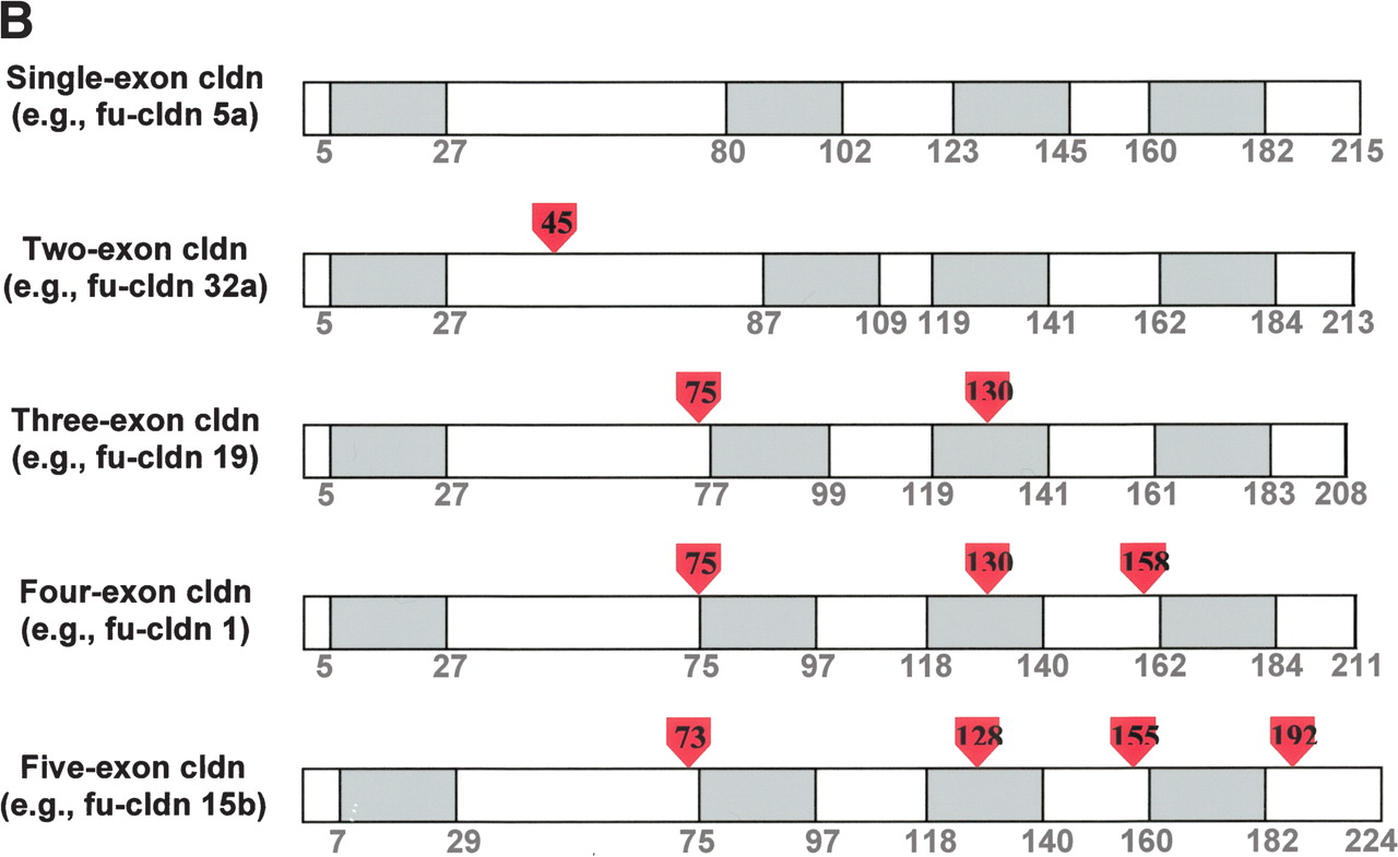
Topology and exon–intron organization of Fugu claudins. (A) Membrane topology of claudins. A diagram of the typical topology of claudins in the lipid bilayer and their location at the tight junction between the apical and lateral plasma membrane of epithelial cells is shown. (B) Exon–intron structure of typical Fugu claudin genes. The approximate location of introns in claudin genes encoded by one, two, three, or four exons is shown based on a typical example. Amino acids are numbered and show the boundaries of the four transmembrane domains (shaded in gray), which are typical for claudins. Intron position is shown by the red arrowheads (the numbers within correspond to the location in relation to the amino acid sequence).


