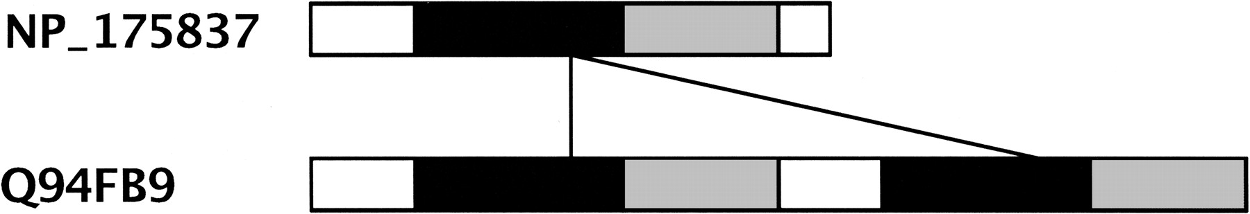
Visualizing Sequence Similarity of Protein Families

(Downloading may take up to 30 seconds. If the slide opens in your browser, select File -> Save As to save it.)

Click on image to view larger version.

Figure 11
Figure 11

The domain structure of two PMP proteins is shown in the figure. The transmembrane domains are colored black, and the nucleotide-binding factors are shown in gray. The two hits between the proteins are shown by black lines.

This Article

  1. Genome Res. 14: 1160-1169

Preprint Server