
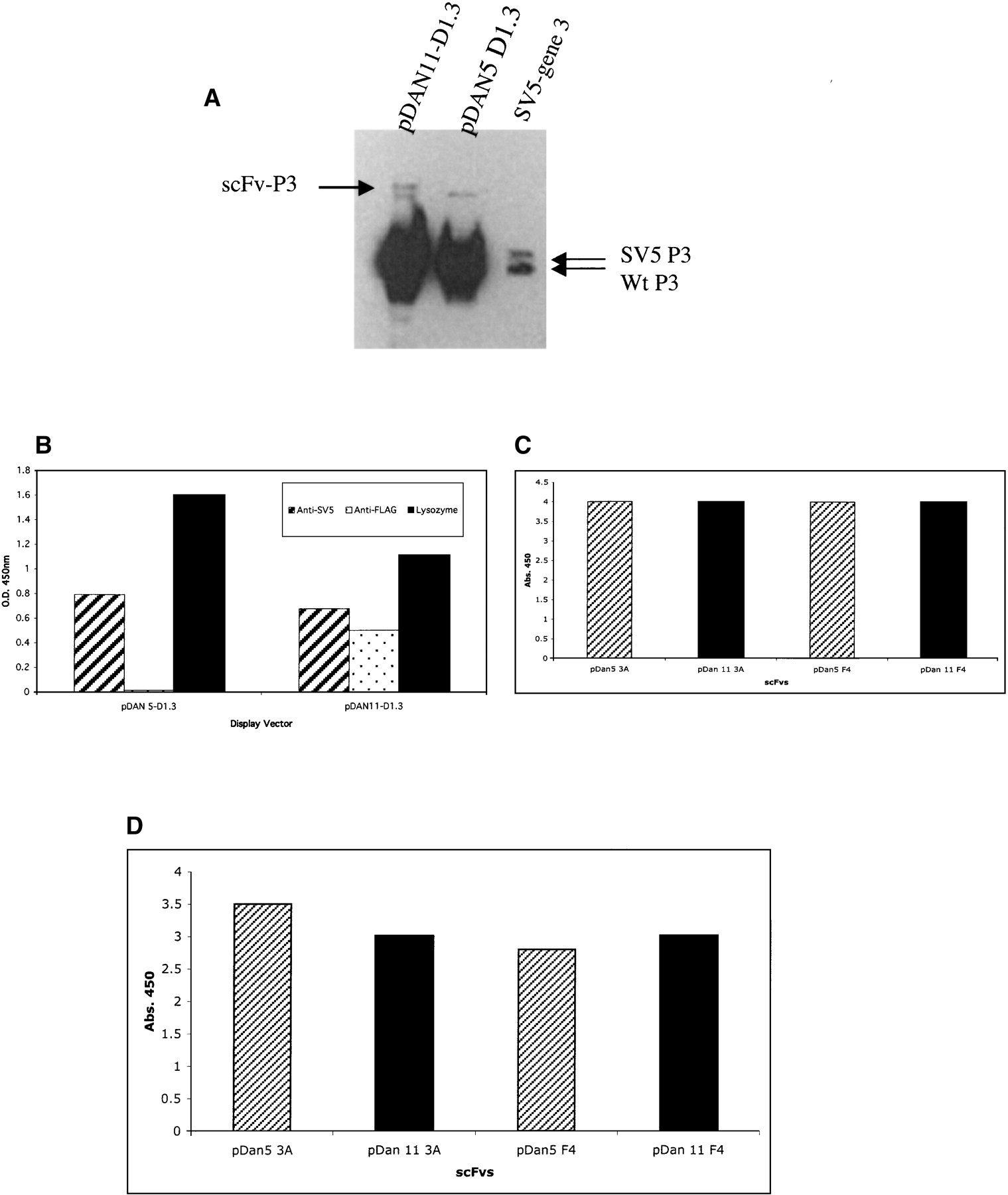
(A) scFv phage display levels. (Lane 1) pDAN11-D1.3; (lane 2) pDAN5-D1.3; (lane 3) a control vector containing only the SV5 tag fused to gene 3 to mark the size of unfused gene 3 protein. Lower arrows denote native gene 3, and upper arrow shows the scFv-gene3 fusion protein. The 2 × 10⁹ phage particles were loaded in lanes 1 and 2, and both the native and fusion forms of gene 3 were visualized using anti-gene3 mAb (1:2000). (B) Phage ELISA signals observed for either pDAN5 D1.3 or pDAN11 D1.3 against anti-SV5 and anti-Flag mAbs, and lysozyme adsorbed to a 96-well plate. Bound phage were determined using anti-M13 HRP (1:5000) conjugate with the background subtracted from the reported values. (C) Phage ELISA. (D) scFv ELISA signals observed for either pDAN5 or pDAN11 3A and F4 scFv against SNV-N adsorbed to a 96-well plate. The bound phage were detected by anti-M13 horseradish peroxidase conjugate (Pharmacia), and the bound scFvs by SV5 anti-tag antibody (Hanke et al. 1992) followed by anti-mouse antibody conjugated to horseradish peroxidase.


