
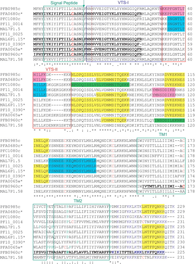
Multiple sequence alignments. The sequences of the Maurer's cleft 2-transmembrane domain proteins were aligned using CLUSTAL W (1.82) (Thompson et al. 1994). (*) Conserved residues. Bold, underlined sequences indicate regions of the proteins that were extended from the originally published gene models (Gardner et al. 2002), on the basis of the complete chromosome sequences that were obtained from PlasmoDB (Bahl et al. 2002). The red-dotted vertical line indicates the boundary between exon 1 and exon 2. The regions covered by peptides identified during the MS/MS analyses are highlighted. The green, magenta, cyan, and yellow color coding stands for peptides unique to a protein, and peptides common to two, three, and more than three proteins, respectively. The green boxes indicate the position of signal peptide and transmembrane domains as predicted by SignalP (Nielsen et al. 1997) and TMHMM (Krogh et al. 2001), respectively. The arrow points at the potential processing site. The red and blue boxes define potential bipartite vacuolar translocation signals (VTSs) as described by Lopez-Estrano et al. (2003).


