
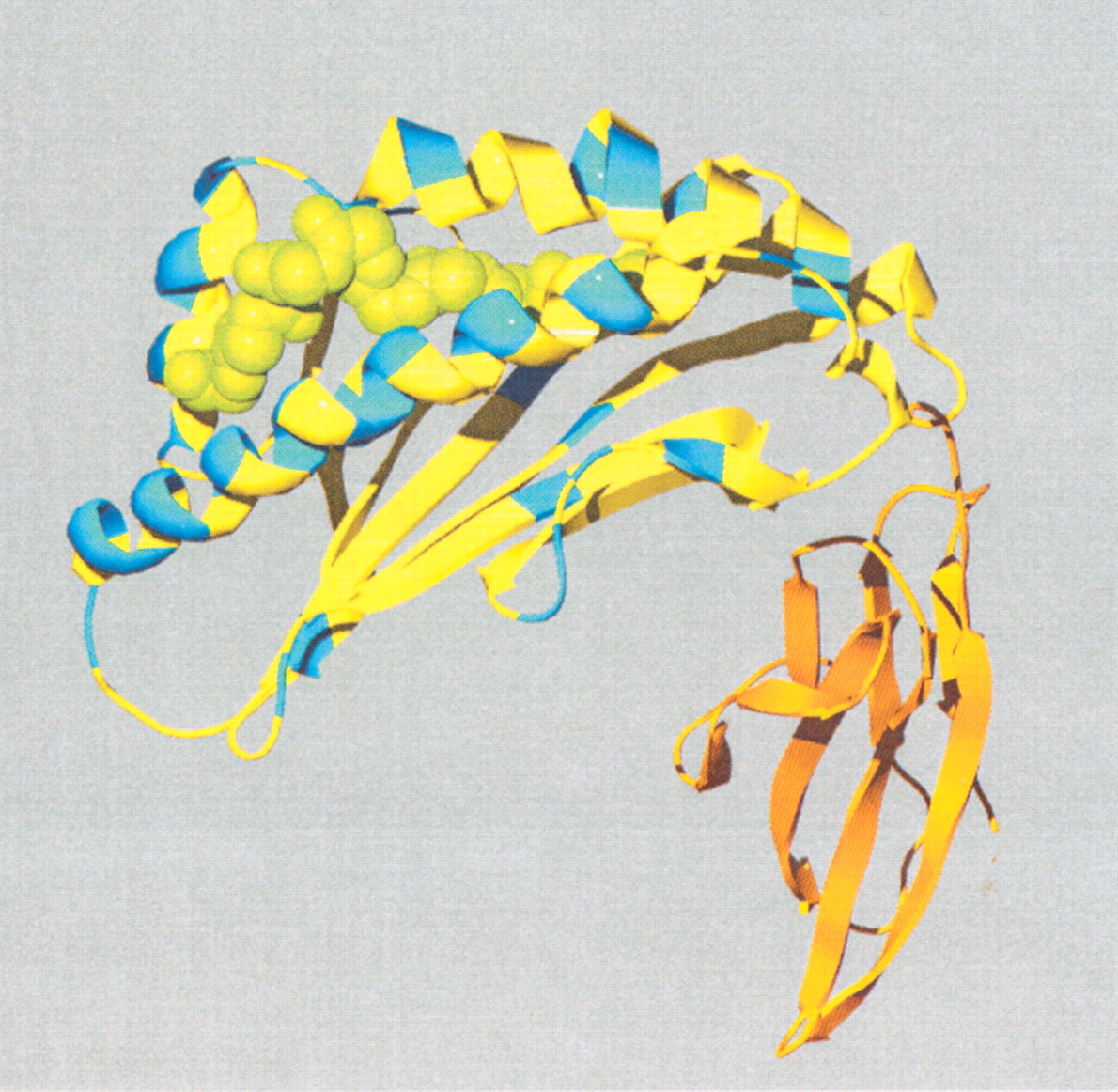
Figure 6
Site-specific KA/KS analysis of MHC-M10 genes. Codons that were predicted to be under positive selection with a posterior probability >0.90 by one codeml model, and >0.5 by at least one other model, are mapped to the crystal structure of mouse MHC class I H-2DD, chain: A in complex with the HIV-1 derived peptide p18–110, shown in green (PDB identifier 1BII (Achour et al. 1998). ω+ sites, shown in blue, are predominantly located along the α-helices of the peptide recognition region. No ω+ sites were mapped to the immunoglobulin domain of the protein, shown here in orange.


