
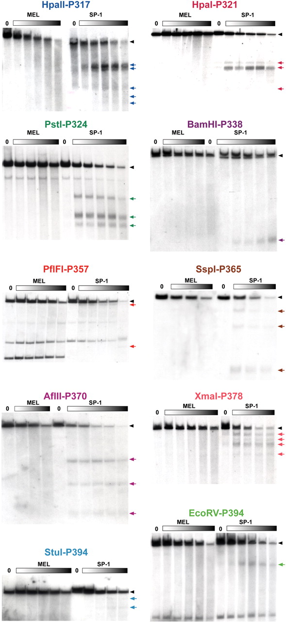
Figure 2.
DNaseI HS mapping of the Sprr locus. Keratinocyte-specific patterns of DNaseI HSs at the conserved regions of the Sprr locus. Nuclei from SP-1 cells and MEL cells were treated with increasing amounts of DNaseI (closed rectangles), and isolated DNAs were digested with restriction enzymes and subjected to Southern blotting with a probe as indicated. The HSs bands are indicated on the right by colored arrows, and their localization in the mouse sequence is shown in Figure 1C. The exact locations of probes and restriction enzyme sites are given in Supplemental Table I.


