
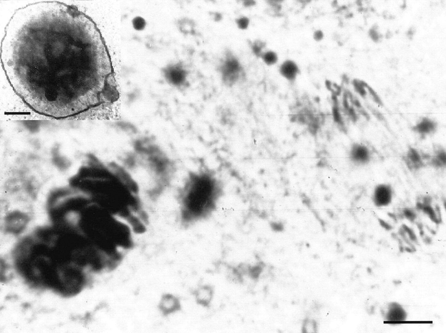
Figure 1.
A microphotograph of the C. kolensis chromosomes in the germline cells (left) and at anaphase of the somatic cells (right) during diminution division. An electron microscope photograph of the granules of eDNA (bar = 5 μ) (top upper corner). A poreless, one-layer membrane surrounds the granule (bar = 1 μ).


