
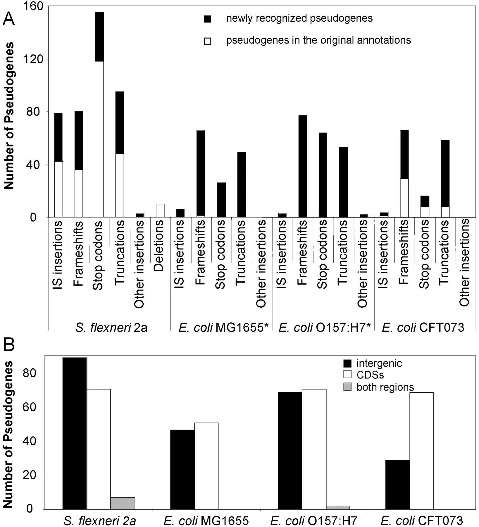
Figure 1.
(A) Numbers and types of mutations forming pseudogenes. Bars include those pseudogenes reported in the original annotation (white) and those recognized subsequently by comparative analyses (black). (The specific mutations labeled as “deletions” in S. flexneri 2a were not provided in the original annotation.) Asterisks denote genomes for which some of the newly recognized pseudogenes include many of those reported in Homma et al. (2002). (B) Locations of newly recognized pseudogenes in relation to annotated CDSs and nonannotated regions.


