
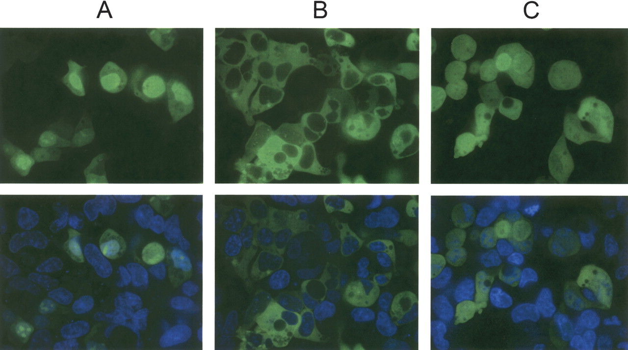
Figure 3
Fluorescent, confocal, live-cell imaging of HEK-293 cells transfected with (A) NLS-eGFP, (B) β-Arrestin-eGFP fusions, or (C) untagged eGFP. Forty-eight hours post-transfection, cells were stained with Hoechst 33342 and imaged confocally at 60× magnification on a Pathway HT system (Atto Bioscience). Pseudo-colored images of a typical Z-stack section are shown. (Top) GFP channel alone; (bottom) Hoechst + GFP.


